Tinidazole
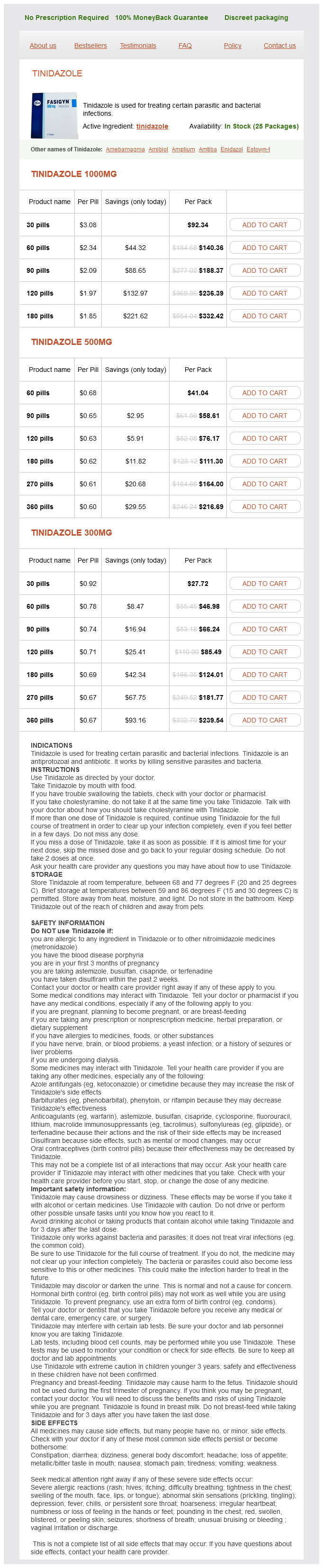
Tinidazole 1000mg
- 30 pills - $92.34
- 60 pills - $140.36
- 90 pills - $188.37
- 120 pills - $236.39
- 180 pills - $332.42
Tinidazole 500mg
- 60 pills - $41.04
- 90 pills - $58.61
- 120 pills - $76.17
- 180 pills - $111.30
- 270 pills - $164.00
- 360 pills - $216.69
Tinidazole 300mg
- 30 pills - $27.72
- 60 pills - $46.98
- 90 pills - $66.24
- 120 pills - $85.49
- 180 pills - $124.01
- 270 pills - $181.77
- 360 pills - $239.54
Failure of closure of the processus vaginalis (inguinal hernia) and abnormal attachment of the gubernacular remnant are common in association with cryptorchidism antibiotics for sinus infection cephalexin 500 mg tinidazole buy free shipping. In their study of 759 patients, Cendron and colleagues identified a persistently patent processus vaginalis ipsilateral to 87% of unilateral and 71% of bilateral undescended testes. Specific notation of the gubernacular position available from this and another large study indicates aberrant attachment lateral to the scrotum in 66% to 75% of cases (Moul and Belman, 1988; Cendron et al, 1993). The processus is patent in 45% to 50% of boys with ascending testes, possibly related to older patient age and/or reduced severity of cryptorchidism in this group (Barthold and Gonzalez, 2003; Barthold et al, 2012; van Brakel et al, 2012). Inguinal hernia is also more common in family members of boys with cryptorchidism (Barthold et al, 2012). Anomalies of the tunica and processus vaginalis in cryptorchidism predispose to development of testicular torsion or clinical hernia, respectively, in rare cases. Torsion of an undescended testis can occur at any age (reviewed by Zilberman et al, 2006) and may be confused with an incarcerated inguinal hernia. The risk of torsion is higher in undescended than in scrotal testes and may be particularly high in children with neuromuscular diseases such as cerebral palsy. Delay in diagnosis is common, and a high index of suspicion is needed to reduce the high risk of testicular loss. Of particular concern is the potential risk for delayed diagnosis with resultant testicular necrosis during the postnatal period when infants are observed for spontaneous testicular descent (Singal et al, 2013). Other Testicular Anomalies Associated with Cryptorchidism Several rare anomalies of testicular development associated with cryptorchidism, each with 100 to 150 reported cases in the literature, include polyorchidism, splenogonadal fusion, and transverse testicular ectopia. Because abdominal cryptorchidism commonly occurs in these cases, laparoscopy is useful in both diagnosis and treatment. Polyorchidism is the presence of a supernumerary testis that is more commonly unilateral and on the left side, with rare cases of bilateral duplication or triplication reported in a comprehensive meta-analysis of the pediatric and adult literature (Bergholz and Wenke, 2009). The cause is unknown, but most authors speculate that this anomaly is related to duplication or division of the genital ridge with or without the wolffian duct as illustrated by Danrad and colleagues (2004). Testes are reported to be scrotal, inguinal, and abdominal in 75%, 20%, and 5% of cases, respectively (Kumar et al, 2008). Affected individuals are frequently asymptomatic, and the polyorchidism is identified at the time of orchidopexy or hernia repair, although a scrotal or inguinal mass and pain with or without torsion may occur and persistent müllerian remnants may coexist. Kumar and colleagues suggest a classification that differentiates between testes that are drained by a vas deferens (type A1- separate epididymis and vas; type A2-separate epididymis; type A3-shared epididymis and vas) and those with no vasal drainage (type B1-epididymis present; type B2-no epididymis or vas). This classification can aid in management decisions that should be based not only on the anatomy of the accessory ducts but also the position, size, and attachments of the testis. Observation and periodic self-examination without surgery should be considered for sonographically normal scrotal testes and orchidopexy for testes that are undescended but with intact ductal drainage (Spranger et al, 2002; Bergholz et al, 2007; Khedis et al, 2008). Occasional cases of testicular tumor have been reported in supernumerary testes, but it is unclear if this is a risk related to polyorchidism per se or to associated cryptorchidism or persistent müllerian duct syndrome (Spranger et al, 2002; Ghose et al, 2007). Splenogonadal fusion is a defect characterized by continuous or discontinuous fibrous union between splenic tissue and the gonad, a condition much more commonly recognized in males (Khairat and Ismail, 2005). In the continuous form (55%) a cord connects the testis to the spleen, whereas in the discontinuous form the splenic tissue is attached to the gonad and not continuous with the main body of the spleen (Ferron and Arce, 2013). Approximately 30% of affected individuals have cryptorchidism, with the majority of cases abdominal and bilateral (59%), and 65% and 26% involving the left and right sides, respectively (Cortes et al, 1996). The continuous form of splenogonadal fusion is more commonly syndromic, associated with limb defects, micrognathia, microglossia, anal atresia, and pulmonary hypoplasia (McPherson et al, 2003) and less commonly with cardiac defects, cleft palate, imperforate anus, and myelomeningocele (Lin et al, 2010). The discontinuous form can manifest as a testicular mass, with distinctive ultrasound findings that include a somewhat hyperechoic mass (relative to testis) that contains multiple hypoechoic nodules and obvious vascularity (Ferron and Arce, 2013). Most cases are found incidentally at the time of orchidopexy or inguinal hernia repair or with scrotal swelling related to illness-related reactive changes within the splenic tissue. Testicular malignancy is reported rarely in association with cryptorchidism and not likely related to the splenic anomaly. Treatment should focus on recognition of the defect at the time of orchidopexy and avoidance of unnecessary orchiectomy. Transverse testicular ectopia may occur as an isolated anomaly in otherwise normal males with cryptorchidism or vanishing testes, or in association with persistent müllerian duct syndrome in 20% to 50% of cases (De Luna et al, 2003; Wuerstle et al, 2007; Thambidorai and Khaleed, 2008). The classic presentation is inguinal hernia with contralateral nonpalpable testis, although both testes may be palpable in the same hemiscrotum.
Tinidazole dosages: 1000 mg, 500 mg, 300 mgTinidazole packs: 30 pills, 60 pills, 90 pills, 120 pills, 180 pills, 270 pills, 360 pills
Bladder stones from children in these developing countries are most often composed of ammonium acid urate antibiotics for uti toddler discount 1000 mg tinidazole mastercard. In contrast, among children from industrialized nations, bladder stones are most often found in children with spinal cord injuries and/or congenital abnormalities such as spina bifida. These children often have undergone augmentation cystoplasty and/or manage their bladders by clean intermittent catheterization. It has been reported that 50% of those children with reconstructed bladders develop bladder stones in their lifetime (Palmer et al, 1993). Urinary stasis, bacterial colonization or infection with urea-splitting organisms, retained mucus, and foreign bodies all can contribute to the formation of bladder stones, most of which are struvite. Transurethral cystolithotripsy is an alternative, although it is not ideal in pediatric patients. In children, a smaller caliber urethra limits effective treatment of large bladder stone burdens. However, percutaneous cystolithotripsy is used worldwide with the advantage of shorter hospital stays, smaller scars, and less indwelling catheter time postoperatively (Al-Marhoon et al, 2009). At the present time, percutaneous cystolithotripsy is the preferred method to treat bladder stones that have formed in reconstructed bladders (Paez et al, 2007). In developing countries, percutaneous lithotripsy has become the first-line treatment modality to address bladder stones in bladders that have not been augmented. For example, Salah and colleagues (2005) reported on their experience with cystolithotripsy in 155 children from Pakistan and Yemen with a mean age of 4. All children were treated safely and successfully using a 26-Fr nephroscope through a 30-Fr sheath placed through a 1-cm suprapubic incision (Salah et al, 2005). Percutaneous cystolithotripsy has been used effectively in infants younger than 1 year to clear bladder stones. Gan and colleagues (2010) reported on their experience using a 16-Fr peel-away sheath with a ureteroscope to treat bladder stones with an average size of 1. Percutaneous cystolithotripsy in children may be performed under ultrasound or fluoroscopic guidance and is most often an outpatient procedure. With either modality, the bladder is first filled to capacity with water or contrast material. The child is placed in Trendelenburg position to minimize the risk of bowel injury during access and tract dilation/formation. An 18-gauge needle is placed into the distended bladder midline, one to two fingerbreadths above the pubic bone. When proper placement is confirmed with return of fluid, a wire is passed through the needle into the bladder. Most often, a tract is established with a balloon dilator to accompany a 30-Fr sheath. A 26-Fr nephroscope is used to extract stones smaller than 1 cm with a rigid stone forceps, or an ultrasonic lithotripter may be used to fragment stones larger than 1 cm. At the conclusion of a percutaneous cystolithotripsy procedure, a Foley catheter is left per urethra or per continent catheterizable stoma for 1 week. Laparoscopy and robotic-assisted laparoscopy have been used successfully in adults for treatment of calculi during the concomitant treatment of ureteropelvic junction obstruction and in the primary treatment of staghorn calculi. Small series using these techniques in children have been described only more recently. Of these cases, four were completed robotically, with one patient having a residual 6-mm lower pole stone and one patient requiring conversion to an open procedure. Urinary stasis, bacterial colonization or infection with urea splitting organisms, retained mucus, and foreign bodies all can contribute to the formation of bladder stones, most of which are struvite. However, despite encouraging results, concern remains regarding safety of endourologic treatment in smaller patients and its subsequent effects on the growing kidney. Prospective studies designed to determine the "preferred" endourologic approach to upper tract calculi in children would be helpful, albeit difficult to conduct. In this regard, individual surgeon experience and comfort level weigh heavily in choosing a treatment modality. Familiarity of pediatric urologists with percutaneous renal access and the full spectrum of endourologic equipment and techniques will continue to facilitate efficacious, minimally invasive approaches to the entire pediatric urinary tract.
Lizzy-Run-Up-The-Hedge (Ground Ivy). Tinidazole.
- What is Ground Ivy?
- Mild lung problems, coughs, arthritis, rheumatism, menstrual (period) problems, diarrhea, hemorrhoids, stomach problems, bladder or kidney stones, wounds or other skin conditions, and other uses.
- Are there safety concerns?
- Dosing considerations for Ground Ivy.
- How does Ground Ivy work?
Source: http://www.rxlist.com/script/main/art.asp?articlekey=96076
Six patients (3%) had minor orthopedic complications including pelvic osteomyelitis in 1 bacteria definition for kids generic tinidazole 300 mg amex, pin site infection in 3, and a pressure sore from immobilization in 1. Dehiscence, which may be precipitated by incomplete mobilization of the pelvic diaphragm and inadequate pelvic immobilization postoperatively, wound infection, abdominal distention, or urinary tube malfunction, necessitates a 4- to 6-month recovery period before a second attempt at closure can be made (Gearhart and Jeffs, 1991a; Gearhart et al, 1993b). Tension-free reclosure with osteotomy and immobilization are important factors in initial and subsequent closures. Unfortunately, the chance of obtaining adequate bladder capacity for bladder neck plasty and eventual continence after multiple closures is markedly diminished (Gearhart et al, 1996a; Novak et al, 2010). Similarly, bladder prolapse is considered a failure and requires bladder reclosure or revision. In a recent series from a single institution of 122 patients who underwent reclosure with a mean follow-up of 14 years, the voided continence rate was 16% (Novak et al, 2010). In a patient with significant bladder prolapse or dehiscence, at the time of secondary closure we combine epispadias repair with bladder, posterior urethral, and abdominal wall closure (Gearhart et al, 1998). The patient is given testosterone enanthate intramuscularly 5 weeks and 2 weeks before surgical repair and undergoes an osteotomy with concomitant bladder, urethral, and abdominal wall closure. Baird and coauthors (2005c) reported on 38 boys undergoing combined exstrophy closure and epispadias repair along with osteotomy; in 30, prior reconstruction had failed. Complications in this group were limited to urethrocutaneous fistulae and urethral strictures. Although the results after reclosure are not as good as those obtained with a successful primary closure, they are respectable and may obviate the need for bladder augmentation. Neourethral stricture is often associated with paraexstrophy skin flap use, pubic suture reaction, erosion, or the use of urethral stents. In a review by Baker and colleagues (1999), posterior urethral obstruction was found in 41 patients. If diversion was used for longer than 6 months, the ultimate fate of the bladder was augmentation. If diversion was used for less than 6 months, most reconstructions were ultimately bowel free. Whether this is a result of ischemic damage to the urethral plate or technical error is unclear. Regardless, because there is a paucity of long-term data from these two repairs, the outlet must be monitored carefully as with all exstrophy patients after primary closure. Ultimately, posterior urethral obstruction after exstrophy closure markedly decreases the success of any repair. This complication presents a significant risk to the upper urinary tract and should be detected early. Although all closed exstrophy bladders have vesicoureteral reflux, upper tract deterioration is the ultimate fate of significant outlet obstruction. At this point the management includes urethral dilation (incision), open urethroplasty, or upper tract diversion. If renal function is compromised, the choice must achieve unquestionable free drainage to allow the upper tracts and kidneys to recover fully. Further bladder neck or urethral reconstruction should not be performed until the posterior urethral stricture is clearly repaired and free drainage has been achieved. Massanyi and colleagues (2012) found that a high number of patients with a vesicocutaneous fistula after primary closure actually have a failed closure. Bladder capacity was the most predictive variable of a successful bladder neck reconstruction after bladder neck injection. In the group of 25 patients with prior bladder neck surgery of whom 16 were partially dry (1 to 3 hours), all were rendered socially continent (dry for longer than 3 hours) after bladder neck injection. Thus, outlet injection after a failed bladder surgery is more successful if a reasonable dry interval has been achieved with open bladder neck surgery. In recent data from Alova and colleagues (2012), bladder neck surgery after prior dextranomer bladder neck injection was not found to be more difficult and success rates were quite acceptable with long-term follow-up. However, repeated dextranomer injections after a prior failure did not result in success. Some successes have been seen with repeated Young-Dees-Leadbetter repair if the bladder neck is patulous, the bladder capacity is adequate, and urodynamic evaluation reveals a stable bladder (Gearhart et al, 1991). In that series only 50% of patients with a failed prior bladder neck reconstruction were candidates for reoperative surgery.
Syndromes
- Pain medicines
- Cancer
- Blood tests
- Bones connect together again, and more surgery is needed
- Scores 6 through 7: Intermediate- (or in the middle) grade cancer. Most prostate cancers fall into this group.
- Malignant hypertension (arteriolar nephrosclerosis)
- Stomach cramps
- Unconsciousness (coma) that continues
- Difficulty talking
- May become severe enough to interfere with activities
With tension on the extra-abdominal testis antibiotics on the pill order tinidazole 1000 mg line, peritoneal attachments overlying the cord can be more easily transected, thus providing addi- tional length. In some cases the testis can only be brought into the upper scrotum; the long-term adequacy of this approach is not clear. Excessive tension on the vessels during placement of the testis should be avoided, because injury or avulsion of the spermatic vessels may occur (Esposito et al, 2002). A key strategy should be preservation of the blood supply between the vas and spermatic artery during dissection so that the Fowler-Stephens procedure can be performed if necessary. Formal closure of the dissected internal ring is not necessary (Handa et al, 2005; Riquelme et al, 2007); indeed, previous experience with open hernia repair suggests that ligation is not needed if the internal ring is dissected (Mohta et al, 2003). No inguinal hernias were identified a mean of 41 to 50 months after laparoscopic orchidopexy in a retrospective series in which formal closure of the internal ring was performed in 54% of cases (Khairi et al, 2013). A contralateral patent processus vaginalis was identified in 9% of boys undergoing laparoscopic orchidopexy in one series, and laparoscopic repair was performed and recommended (Palmer and Rastinehad, 2008). However, the necessity for this approach in preventing clinical hernia formation is questionable based on studies of boys undergoing laparoscopic or open hernia repair (Schier, 2007). For testes that are not near (variably defined as 2 to 4 cm above) the internal inguinal ring, transection of the spermatic vessels as originally described by Fowler and Stephens may be necessary (Fowler and Stephens, 1959); a long-looping vas facilitates but is not required for testicular mobilization to the scrotum. The FowlerStephens procedure is now typically performed laparoscopically with spermatic vessel clipping (Bloom, 1991) followed by laparoscopic or open testicular mobilization in the same setting, or in a staged approach 6 months later. The peritoneum should be left intact over the vasal vessels, and the gubernacular vessels should be left intact if possible. This group subsequently studied the effect of low versus high transection of the vessels in prepubertal rats and showed a reduction in adult testicular sperm numbers that was similar in both groups (Srinivas et al, 2005). In human studies, testicular biopsies before and after spermatic vessel transection also showed a reduction in germ cell count, a finding that was significant in younger boys (Thorup et al, 1999; Rosito et al, 2004). In general, the preferred approach is avoidance of spermatic vessel transection whenever possible; the available data suggest this is possible in the majority of cases of abdominal orchidopexy. In rare cases, particularly if the testis is retrovesical, the vas is too short to allow scrotal placement of the testis, and orchiectomy is ultimately required (Perovic and Janic, 1997). The success rates for laparoscopic procedures as shown in Table 148-1 appear to compare favorably with the corresponding 74%, 63%, and 77% overall success rates for open surgical and one-stage and two-stage Fowler-Stephens procedures, respectively (Docimo, 1995). The available data suggest that a primary procedure is more consistently successful (>90% in most series) than a FowlerStephens approach (variable success of 60% to 97%). In directly comparing the results of 156 abdominal orchidopexies at a single institution, Stec and colleagues observed significantly better results for a primary open (89% success) or laparoscopic (97%) approach than for one-stage (63%) or two-stage (68%) Fowler-Stephens procedures (Stec et al, 2009). Recent meta-analyses and/or systematic reviews of surgical treatment of abdominal testes (Elyas et al, 2010; Guo et al, 2011; Penson et al, 2013; Kolon et al, 2014) are primarily low-quality retrospective series with few, if any, adequately powered prospective controlled studies. Pooled success rates for primary onestage Fowler-Stephens and two-stage Fowler-Stephens procedures are approximately 95%, 80%, and 85%, respectively. The available evidence suggests no clear difference in efficacy between open and laparoscopic procedures. Overall success refers to the frequency of nonatrophic testes in satisfactory scrotal position according to variably detailed criteria used by the authors. Despite their limitations, the available data seem to suggest that primary orchidopexy without transection of the spermatic vessels is preferable whenever possible. Some authors recommend that ultrasound be used to confirm testicular viability postoperatively (Esposito et al, 2002). Other complications of laparoscopic orchidopexy are rare and potentially include bladder or vascular injury, hypercapnia, and delayed small bowel obstruction (Esposito et al, 2003; Hsieh et al, 2009). Laparoscopic techniques may be applicable in unusual cases, including bilateral orchidopexy, abdominal wall defects, polyorchidism, splenogonadal fusion, and transverse testicular ectopia with or without persistent müllerian ducts. Some surgeons have considered microvascular orchidopexy to be a preferred approach to the solitary abdominal testis, particularly with historical success rates of 88% as compared with lower rates for open procedures (Docimo, 1995). At a center with substantial experience using the microvascular approach, long-term success rates of 96% for standard and 88% for laparoscopically assisted autotransplantation were reported (Bukowski et al, 1995; Tackett et al, 2002). The advantage of this approach is preservation of the spermatic vessels, at the cost of longer operative time and requirements for an experienced microvascular surgeon and hospital stay.
Usage: t.i.d.
In their treatment protocol virus 2014 symptoms cheap tinidazole 300 mg buy, asymptomatic remnants are managed with physical and sonographic examination. They found that spontaneous resolution with nonoperative management is likely with remnants in patients younger than 6 months. However, if symptoms persist or the remnant fails to resolve after age 6 months, they recommend excision. Vesicourachal diverticulum (3% to 5%) Patent Urachus Patent urachus is explained by nondescent of the bladder or, more commonly, failure of the epithelial-lined urachal canal to obliterate (Gearhart, 2002). Bladder obstruction during fetal development has been blamed for the urachus remaining tubular. The fact that urachal patency is often absent with severely obstructed bladders in utero, however, casts doubt on this theory. Additionally, only up to 14% of patients with a patent urachus have postnatal confirmation of in utero bladder obstruction (Schrenck and Campbell, 1972; Mesrobian et al, 1997). It seems possible that obliteration of the urachus may be independent from the level of bladder distention. Therefore retubularization, rather than primary patency, might be the cause for urinary drainage from the umbilicus. Chapter138 BladderAnomaliesinChildren 3177 Patent urachus would manifest as a Meckel diverticulum connected to the umbilicus. These structures can be very difficult to differentiate from an umbilical-urachus sinus because no connection to the bladder or bowel can be seen on sinugram. However, the surgical approach to both anomalies requires the complete excision of all tissue. Unlike urachal structures, omphalomesenteric remnants can show gastric or small bowel mucosa on histologic examination. However, the fluid-filled cyst can drain through the umbilicus or into the bladder intermittently. Urachal cysts are found more commonly in the distal part of the urachus and manifest more commonly in adults than in infants or children (Cilento et al, 1998). These cells can become infected; Staphylococcus aureus has been identified as the most common organism (Mesrobian et al, 1997). Once infected, urachal cysts can manifest as umbilical abscess formation or bladder infections. Additional symptoms include localized lower abdominal pain, voiding symptoms, or even a painful and palpable mass. The diagnosis is confirmed by ultrasound, demonstrating the localized cyst between the anterior abdominal wall and the peritoneum. If unrecognized, the infected cyst can perforate into the bladder (Maruschke et al, 2003) or peritoneal cavity. This can cause peritonitis and formation of an enteric fistula (Ohgaki et al, 2003; Quek et al, 2003). Treatment consists of draining the infected cyst, followed by complete excision of the urachal remnant structures. The rectus fascia is incised in a longitudinal fashion, muscles spread apart, and the dome of the distended bladder identified. The dissection continues extraperitoneally toward the umbilicus until the previously incised umbilical portion is free and can be pulled into the surgical field. A bladder cuff including the urachal insertion is marked and excised using electrocautery. This can be done even in children younger than 6 months of age (Fahlenkamp et al, 1995; Cadeddu et al, 2000; Khurana and Borzi, 2002). Khurana and Borzi (2002) described their laparoscopic experience with 4 children between 5 months and 10 years and found the procedure to be safe in children of all ages. Technically, they suggested a three-port approach, with the camera port in the midline between the umbilicus and the xiphoid and two working ports on either side in the upper quadrant. Early in their series they inserted the camera port at the umbilicus with working ports into the right and left upper abdomen. Later they preferred to place the camera into the left lower abdominal wall, with 2-mm working ports at the left lower and upper abdomen for better visualization of the urachus at the umbilicus. They reported no intraoperative or postoperative complications, no recurrences, and a median operating time of 35 minutes.
References
- Duffy JF, Willson HJ, Wang W, et al: Healthy older adults better tolerate sleep deprivation than young adults, J Am Geriatr Soc 57:1245-1251, 2009.
- Ortega AE, Hunter JG, Peters JH, et al. A prospective randomized comparison of laparoscopic appendectomy with open appendectomy. Am J Surg 1995;169:208-12.
- Mauermann WJ, Nemergut EC: The anesthesiologist's role in the prevention of surgical site infections, Anesthesiology 105(2):413-421, quiz 439-440, 2006.
- Gutierrez C, et al: An experimental study on the effects of ethanol and folic acid deficiency, alone or in combination, on pregnant Swiss mice, Pathology 39(5):495-503, 2007.
- Sternberg CN, Hawkins RE, Wagstaff J, et al: A randomised, double-blind phase III study of pazopanib in patients with advanced and/or metastatic renal cell carcinoma: final overall survival results and safety update, Eur J Cancer 49:1287n1296, 2013.