Sildenafilo
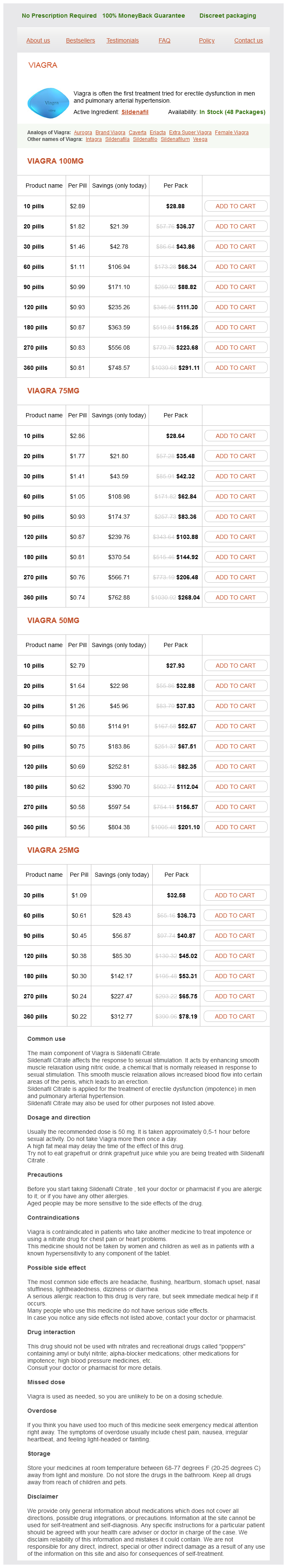
Viagra 100mg
- 10 pills - $28.88
- 20 pills - $36.37
- 30 pills - $43.86
- 60 pills - $66.34
- 90 pills - $88.82
- 120 pills - $111.30
- 180 pills - $156.25
- 270 pills - $223.68
- 360 pills - $291.11
Viagra 75mg
- 10 pills - $28.64
- 20 pills - $35.48
- 30 pills - $42.32
- 60 pills - $62.84
- 90 pills - $83.36
- 120 pills - $103.88
- 180 pills - $144.92
- 270 pills - $206.48
- 360 pills - $268.04
Viagra 50mg
- 10 pills - $27.93
- 20 pills - $32.88
- 30 pills - $37.83
- 60 pills - $52.67
- 90 pills - $67.51
- 120 pills - $82.35
- 180 pills - $112.04
- 270 pills - $156.57
- 360 pills - $201.10
Viagra 25mg
- 30 pills - $32.58
- 60 pills - $36.73
- 90 pills - $40.87
- 120 pills - $45.02
- 180 pills - $53.31
- 270 pills - $65.75
- 360 pills - $78.19
The region between the inner and outer hair cells is occupied by pillar cells erectile dysfunction forums cheap 75 mg sildenafilo, which assist in the formation of the inner tunnel (of Corti). Nerve fibers lead to the spiral ganglion (housing pseudounipolar cells) in the modiolus. Membranous Labyrinth the membranous labyrinth is composed of the utricle, the saccule, and the three semicircular canals. Utricle and Saccule the utricle and saccule are both filled with endolymph and house maculae. Each macula is composed of simple columnar epithelium composed of two cell types, neuroepithelial hair cells and supporting cells. The free surface of the macula displays the otolithic membrane, housing small particles called otoliths. Semicircular Canals the three semicircular canals are oriented perpendicular to each other. The ampulla of each canal houses a crista, a structure similar to a macula, composed of neuroepithelial hair cells and supporting cells. A gelatinous cupula is located at the free surface of the crista, but it contains no otoliths. Appendix the light microscopic study of cells, tissues, and organs requires that the material to be examined be sectioned thinly enough to permit light to penetrate it and that the light be sufficient to be collected by the lenses of the microscope and to reach the retina of the examiner. Moreover, the tissue has to maintain its natural, living characteristics; otherwise, the viewer will have a distorted picture of the tissue. Over the years, numerous procedures were developed and refined to make sure that there is a close resemblance between the image under the microscope and the properties of the tissue while it was in its living state. These procedures include fixation, dehydration, clearing, embedding, sectioning, mounting, staining, and the affixing of a coverslip over the section. For light microscopy, the fixative of choice is neutral buffered formalin, although many other fixatives are commonly used. Tissues embedded in paraffin are usually sectioned 5 to 10 m in thickness, whereas those embedded in plastic are sectioned much thinner (0. Because many of the stains used are water miscible, the sections must be deparaffinized and rehydrated before they can be stained. Terminology of Staining Frequently, when staining histological sections, a principal stain is used in conjunction with a counterstain, a contrasting color that stains those components of the tissue that are not stained well with the principal stain. Usually, the stains are either acidic (anionic) or basic (cationic) and are attracted to those components of the cell or tissue that are basic or acidic, respectively. Therefore, the acidic components of the cell, such as nucleic acids, attract the basic stains and are said to be basophilic. Those components of the cell whose pH is greater than 7, such as many cytoplasmic proteins, attract acidic stains and are said to be acidophilic. Common Stains Used in Histology Although a great number of histologic and histopathologic stains have been developed, only the most commonly used stains are listed in this Appendix. Hematoxylin is a basic stain that dyes nuclei, nucleoli, and ribosomes blue to purple in color. Eosin stains basic components of the cell, including myofilaments of muscle, pink to light red in color. Additionally, extracellular matrix proteins, such as collagen, are also stained pink to light red. Weigert Method for Elastic Fibers and Elastic van Gieson Stain Weigert method and van Gieson stain for elastic fibers are both used commonly to stain elastic fibers. Since nuclei also stain dark gray to black, the fibroblasts present among the elastic fibers are very difficult to see. Some cells, such as diffuse neuroepithelial cells, also stain with silver stains and were called argentaffin or argyrophilic cells. Wright Stain Wright stain and the related Giemsa stain are designed specifically for staining cells of blood. It stains erythrocytes salmon pink; nuclei of leukocytes and granules of platelets stain dark blue to purple, whereas the specific granules of eosinophils stain salmon pink, and those of basophils stain dark blue to black. Thus, basement membranes stain pinkish red, whereas mucins of goblet cells and of mucous salivary glands stain a deep red to magenta.
Sildenafilo dosages: 100 mg, 75 mg, 50 mg, 25 mgSildenafilo packs: 10 pills, 20 pills, 30 pills, 60 pills, 90 pills, 120 pills, 180 pills, 270 pills, 360 pills
This gradient gradually increases from about 300 mOsm/L in the interstitium of the outer medulla to as much as 1 impotence with beta blockers best purchase for sildenafilo,200 mOsm/L at the renal papilla. Aldosterone prompts additional resorption of sodium and chloride from the ultrafiltrate located in the distal convoluted tubule. Ascending Thin Limb the removal of the fluid and salts transported into the renal interstitium by the uriniferous tubules. The third muscle layer, the outermost longitudinal layer, appears in the lower one-third of the ureter. Folding occurs at the interplaque regions, whereas the thickened plaque regions present vesicular profiles, which probably become unfolded as urine accumulates in the bladder. The lamina propria of both sexes contains mucous glands of Littré and intraepithelial glands, which lubricate the lining of the urethra, facilitating the passage of urine to the outside. Concentration of Urine in the Collecting Tubule the ultrafiltrate that enters the collecting tubule is hypoosmotic. As it passes down the collecting tubule, it is subject to the increasing osmotic gradient of the renal interstitium. The high interstitial osmolarity of this region is attributed to the urea concentration. Role of the Vasa Recta in Urine Concentration (Countercurrent Exchange System) the vasa recta assists in the maintenance of the osmotic concentration gradient of the renal medulla, since these capillary loops are completely permeable to salts and water. Normal urine is either colorless or has a yellow color if the urine is concentrated. Similarly, dilute urine has very little odor, whereas concentrated urine has a pungent smell. If the color of urine is reddish, the individual may have porphyria or there is fresh blood in the urine; if the color is brown, the possibility is that breakdown byproducts of damaged muscle or breakdown by-products of hemoglobin are in the urine. Black discoloration could be due to the presence of melanin pigment in the urine, whereas cloudy urine could be an indication of the presence of acidic crystals or the presence of pus derived from urinary tract infection. Additionally, certain medications can discolor the urine, and the patient should be warned in advance about the color change. Changes in the odor of urine can be due to diabetes that is not being controlled (a sweet odor); fetid odor could indicate the presence of a urinary tract infection; and a musty odor of urine in a young patient may suggest phenylketonuria. The excess fluid loss in the formation of copious quantities of dilute urine results in polydipsia (excessive thirst) and dehydration. Diabetic Glomerulosclerosis Diabetes mellitus causes vascular pathologies that involve blood vessels throughout the body, including those of the glomerular capillary network where synthesis of the basement membrane components increases to such an extent that it interferes with normal filtration. Additionally, hypercellularity of the mesangial cell population also interferes with the function of the normal Tubular Necrosis Tubular necrosis may result in acute renal failure. Cells of the renal tubules die either by being poisoned due to exposure to toxic chemicals, such as mercury or carbon tetrachloride, or die because of severe cardiovascular shock that reduces blood flow to the kidneys. If the basal laminae remain intact, epithelial cell division may be able to repair the damage in less than 3 weeks. Acute Glomerulonephritis Acute glomerulonephritis is usually the result of a localized beta streptococcal infection in a region of the body other than the kidney. Plasma cells secrete antibodies that complex with streptococcal antigens, forming an insoluble antigen-antibody complex that is filtered by the basal lamina between the podocytes and the endothelial cells of the glomerulus. As the immune complex builds up in the glomerular basal lamina, the epithelial cells and mesangial cells proliferate. Additionally, leukocytes accumulate in the glomerulus, congesting and blocking it. Moreover, pharmacologic agents released at the site of damage cause the glomerulus to become leaky, and proteins, platelets, and erythrocytes may enter the glomerular filtrate. Usually after the acute inflammation abates, the glomeruli repair themselves and the normal kidney function returns. Occasionally, however, the damage is extensive and kidney function becomes permanently impaired. This figure is from the kidney of a patient with end-stage renal disease as a result of diabetes mellitus. Note that the glomerular capillaries are engorged with blood, the intraglomerular cell population is increased, and the glomerular basement membrane displays evidence of being thickened.
L-3-hydroxy-4-(trimethylammonium)-butyrate (L-Carnitine). Sildenafilo.
- Improving low birth weight.
- Dosing considerations for L-carnitine.
- Are there any interactions with medications?
- Preventing side effects caused by valproic acid (Depacon, Depakene, Depakote, VPA), a seizure medication.
- Are there safety concerns?
Source: http://www.rxlist.com/script/main/art.asp?articlekey=96985
The central aminocyclitol for streptomycin is streptidine impotence depression purchase sildenafilo us, whereas for all other current aminoglycosides, it is 2-deoxystreptamine. The aminocyclitol ring is numbered counterclockwise, and the linked sugar molecules clockwise. Both drugs contain a distinctive pentose linkage as well as linkage to two hexose sugars. Among the commonly used aminoglycosides, neomycin contains the largest number of free amino groups (six). Kanamycin, tobramycin, amikacin, arbekacin, and dibekacin comprise the kanamycin family (Table 25-2). Amikacin is kanamycin A with the semisynthetic addition of 2-hydroxy-4-aminobutyric acid to the amino group at position 1 of the aminocyclitol. Arbekacin and dibekacin are available for clinical use outside of the United States. Gentamicin is a mixture of C1, C1a, and the enantiomers C2 and C2a elaborated by Micromonospora spp. Sisomicin is the dihydro analogue of gentamicin C1a, and netilmicin is derived from sisomicin by the addition of an ethyl group to the amino group at position 1 of the aminocyclitol. Resolving the Antibiotic Paradox: Progress in Understanding Drug Resistance and Development of New Antibiotics. The sites of action of four inactivating enzymes are shown, including three acetyltransferases-Aac(3), Aac(2), and Aac(6)-and one adenyltransferase, Ant(2). The overall positive charge contributes to both antimicrobial activity and toxicity. Antibacterial activity is enhanced in media with an alkaline pH and reduced in media with an acidic pH. In vitro, gentamicin and tobramycin are inactivated with greater ease than netilmicin, amikacin, or isepamicin. Perhaps because their dosage is in grams rather than milligrams, the antipseudomonal penicillins (carbenicillin, ticarcillin, piperacillin, mezlocillin, and azlocillin) are the -lactams most susceptible to the reaction. In general, penicillins and aminoglycosides should not be mixed in the same solution before infusion, although a recent product reformulation does allow piperacillin/tazobactam co-administration with gentamicin at specific doses. If patients with renal failure are given an aminoglycoside and an antipseudomonal penicillin concomitantly, there is a 10% to 20% reduction in the serum aminoglycoside concentration compared with the levels observed when each drug is administered alone. The respective nucleotide sequence differences between bacterial and human ribosomal subunits leads to lower affinity of aminoglycosides for the latter. In eukaryotic cytosolic ribosomes, adenosine is replaced by a guanosine within the A site, with a concomitant reduction in binding of aminoglycosides. The double-adenosine nucleotides in prokaryotic ribosomes create an internal bulge and a larger groove that allows access to the ribosomal binding site. Puglisi, Director, Stanford Magnetic Resonance Laboratory, Stanford University School of Medicine. Initial electrostatic binding of aminoglycosides to the cell surface is followed by two energy-dependent uptake phases and binding to ribosomes. Chloramphenicol and other drugs that inhibit protein synthesis are bacteriostatic and not bactericidal, which suggests additional unidentified mechanisms of bactericidal activity. Recent work suggests that only bactericidal drugs stimulate hydroxyl radical formation in bacteria as a function of metabolism-related depletion of reduced nicotinamide adenine dinucleotide, destabilization of iron-sulfur clusters, and stimulation of the Fenton reaction. It is suggested that the high intracellular concentrations result from aminoglycoside closure of voltage-gated channels, with subsequent trapping of drug. Some 1800 genetic diseases arise, in part, from base pair insertions, deletions, or substitutions that produce premature stop codons, resulting in production of incorrectly truncated, nonfunctional proteins. Aminoglycosides can suppress premature stop codons and restore physiologically active amounts of functional protein. Biofilm formation is a concern in the treatment of chronic infections, especially those with foreign bodies. Low-level aminoglycoside resistance attributed to impaired cell wall permeability may be the result of drug efflux mechanisms. Multidrug (including aminoglycoside) efflux pumps are adenosine triphosphatedependent active pumps recently recognized as major contributors to antibiotic resistance. A subpopulation results from small colony variants with deficient energy-dependent uptake of aminoglycosides and may result in clinical treatment failure. The enzymatically modified drugs bind poorly to ribosomes and result in high levels of resistance.
Syndromes
- Permanent changes in the shape of the nail
- Avoid colored or perfumed toilet tissue and bubble bath.
- Bleeding
- Get plenty of calcium and vitamin D in food or supplements.
- Having to sit down to urinate
- Breathing support (possibly artificial respiration)
- Polymyositis
- Headache
The upper third of the esophageal muscularis externa is all skeletal muscle; the middle third is a mixture of skeletal and smooth muscle; and the lower third is all smooth muscle erectile dysfunction fruit sildenafilo 100 mg order online. The gastric mucosa is lined by a simple columnar epithelium whose surface lining cells (not goblet cells) produce a mucous substance that coats and protects the stomach lining from the low pH environment and from autodigestion. The lamina propria of the stomach houses gastric glands; depending on the region of the stomach, these are cardiac, fundic, or pyloric (see Graphic 14-1). The luminal surface, lined by a simple columnar epithelium (surface lining cells), displays foveolae (gastric pits), whose base is perforated by several gastric glands of the lamina propria. Parietal cells live for approximately 200 days before being replaced by stem cells. Parietal cells also secrete gastric intrinsic factor, a glycoprotein that binds to and forms a complex with vitamin B12 in the gastric lumen. When this complex reaches the ileum, it binds to specific receptors on the surface absorptive cells, and the vitamin becomes absorbed (see Table 14-2). They are located in the neck of gastric glands, and they manufacture soluble mucus that becomes part of and lubricates chyme. Surface lining cells live for about 3 to 5 days and manufacture visible mucus that adheres to the lining of the stomach, protecting it from autodigestion. They produce hormones such as gastrin, somatostatin, secretin, and cholecystokinin. Regenerative cells, located mainly in the neck and isthmus, replace the epithelial lining of the stomach and the cells of the glands. Chief cells, located in the base of the fundic glands, live for about 60 to 90 days. Small Intestine the small intestine is composed of the duodenum, jejunum, and ileum. The mucosa of all three regions displays villi (singular: villus), extensions of the lamina propria, covered by a simple columnar type of epithelium. Their tips have a thick coat of glycocalyx, rich in disaccharidases and dipeptidases. These cells function in absorption of sugars, amino acids, fatty acids, monoglycerides, electrolytes, water, and many other substances. These epithelial cells also participate in the immune defense of the body by manufacturing secretory protein, which binds to the J protein component of the antibody and protects immunoglobulin A (IgA) as it traverses the epithelial cell and enters the intestinal lumen. Long-chained lipids, in the form of chylomicrons, are delivered to the lacteals, blindly ending lymphatic channels of the villus. Simple tubular glands of the mucosa, the crypts of Lieberkühn, open into the intervillar spaces. Large Intestine the large intestine is subdivided into the cecum, the colon (ascending, transverse, descending, and sigmoid), the rectum, the anal canal, and the appendix (see Graphic 14-2). The large intestine possesses no villi but does house crypts of Lieberkühn in its lamina propria. The large intestine functions in the absorption of the remaining amino acids, lipids, and carbohydrates, as well as fluids, electrolytes, and certain vitamins, and it also is responsible for the compaction of feces. The cells then release the glucose and galactose into the lamina propria, where these sugars enter the circulatory system for transport to the liver. Since only a thin, simple columnar epithelium separates the richly vascularized connective tissue from this threatening milieu, the lamina propria of the intestines is well endowed with lymphoid elements. These include · scattered cells (B cells, T cells, plasma cells, mast cells, macrophages, etc. Regions where lymphatic nodules come in contact with the epithelial lining of the intestines display flattened cells that form the interface between the lumen and the lymphatic nodule. These are M cells (microfold cells), which phagocytose antigens and transport them (without breaking them down into epitopes), via clathrincoated vesicles, to the basal aspect of the cell. The antigens are released into the lamina propria for uptake by antigen-presenting cells and dendritic cells. It is interesting to note that the lamina propria of the colon has no lymph vessels, thus, generally, cancers of the colon metastasize at a much slower rate than do other cancers of the digestive tract.
Usage: p.r.n.
Antibacterial action of "broad-spectrum" penicillins impotence homeopathy treatment discount sildenafilo 75 mg line, cephalosporins and other antibiotics against gram-negative bacilli isolated from bacteremic patients. In vitro activity of apalcillin compared with that of other new penicillins and antiPseudomonas cephalosporins. Reassessment of susceptibility test interpretive criteria for ticarcillin and ticarcillin-clavulanic acid. Antimicrobial activity and spectrum investigation of eight broadspectrum -lactam drugs: a 1997 surveillance trial in 102 medical centers in the United States. Comparative in vitro activities of piperacillin-tazobactam and ticarcillin-clavulanate. Susceptibility of "enterobacteria" to penicillins, cephalosporins, lincomycins, erythromycin, and rifampin. Nafcillin: antibacterial action in vitro and absorption and excretion in normal young men. Comparative study of piperacillin, ticarcillin, and carbenicillin pharmacokinetics. Pharmacokinetics of amoxicillin: dosage nomogram for patients with impaired renal function. Pharmacokinetics of flucloxacillin and cloxacillin in healthy subjects and patients on chronic intermittent haemodialysis. Incidence of -lactam-induced delayed hypersensitivity and neutropenia during treatment of infective endocarditis. Incidence and risk factors of oral antibiotic-associated diarrhea in an outpatient pediatric population. Comparative in vitro potency of amoxicillin-clavulanic acid and four oral agents against recent North American clinical isolates from a global surveillance study. Amoxicillin/ clavulanic acid: a review of its antibacterial activity, pharmacokinetics and therapeutic use. Antimicrobial activities of piperacillin-tazobactam against Haemophilus influenzae isolates, including -lactamase-negative ampicillin-resistant and -lactamase-positive amoxicillinclavulanate-resistant isolates, and mutations in their quinolone resistance-determining regions. National survey on the susceptibility of Bacteroides fragilis group: report and analysis of trends for 1997-2000. Andes Although the discovery of the cephalosporin antibiotic class was reported in 1945, it took almost 2 decades for this class to achieve clinical utility. Giuseppe Brotzu is widely credited for discovery of the broad-spectrum inhibitory effects of sewage outflow in Sardinia, Italy. He also demonstrated the in vivo activity of these culture filtrates in animal infection models and in several patients. The filtrate was administered both locally by injection into skin abscesses and systemically for the treatment of brucellosis and typhoid fever. A decade after the initial discovery, the cephalosporin substances were isolated and identified as fermentation products of the mold. Each of the products possessed antimicrobial activity but only cephalosporin C demonstrated activity against both gram-negative and gram-positive bacteria. In addition, it had advantageous stability in the presence of acid and penicillinases. The first cephalosporin pharmaceutical, cephalothin, was introduced for clinical use in 1964. There are more than 20 cephalosporin antibiotics in use today (17 in the United States). The cephalosporin class is among the most widely prescribed antimicrobial classes because of its broad spectrum of activity, low toxicity, ease of administration, and favorable pharmacokinetic profile. Changes in the R2 constituent may influence the ability of the compound to reach certain infection sites, such as the central nervous system, or may simply prolong the elimination half-life of the drug. The predominant changes at R1 (position C7) include the addition of an acyl side chain and the substitution of the hydrogen with a methoxy group.
References
- Caplan LR. Migraine and vertebrobasilar ischemia. Neurology 1991;41(1):55-61.
- Cordova MJ, Giese-Davis J, Golant M, et al. Mood disturbance in community cancer support groups. The role of emotional suppression and fighting spirit. J Psychosom Res 2003;55(5):461-467.
- Fiore MC, Jorenby DE, Schensky AE, et al. Smoking status as the new vital sign: effect on assessment and intervention in patients who smoke. Mayo Clin Proc 1995;70(3):209-213.
- Ball CG, Kirkpatrick AW, Laupland KB, et al. Incidence, risk factors, and outcomes for occult pneumothoraces in victims of major trauma. J Trauma. 2005;59(4):917-924; discussion 924-915.