Quetiapine
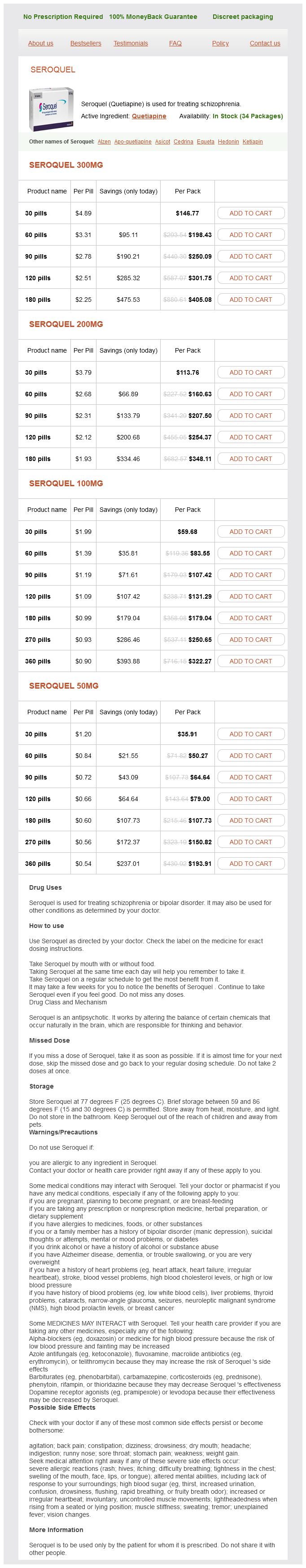
Seroquel 300mg
- 30 pills - $146.77
- 60 pills - $198.43
- 90 pills - $250.09
- 120 pills - $301.75
- 180 pills - $405.08
Seroquel 200mg
- 30 pills - $113.76
- 60 pills - $160.63
- 90 pills - $207.50
- 120 pills - $254.37
- 180 pills - $348.11
Seroquel 100mg
- 30 pills - $59.68
- 60 pills - $83.55
- 90 pills - $107.42
- 120 pills - $131.29
- 180 pills - $179.04
- 270 pills - $250.65
- 360 pills - $322.27
Seroquel 50mg
- 30 pills - $35.91
- 60 pills - $50.27
- 90 pills - $64.64
- 120 pills - $79.00
- 180 pills - $107.73
- 270 pills - $150.82
- 360 pills - $193.91
Other subtle signs include quadriceps wasting treatment yeast infection home buy quetiapine 100 mg with visa, fluid in the knee, tenderness under the edge of the patella and crepitus on moving the knee. Patellofemoral pain is elicited by pressing the patella against the femur and asking the patient to contract the quadriceps first with central pressure, then compressing the medial facet and then the lateral. If, in addition, the apprehension test is positive, this suggests previous subluxation or dislocation. Patellar tracking can be observed with the patient seated on the edge of the couch, flexing and extending the knee against resistance; in some cases subluxation is obvious. With the patient sitting or lying supine, patellar alignment can be gauged by measuring the quadriceps angle, or Q-angle the angle subtended by the line of quadriceps pull (a line running from the anterior superior iliac spine to the middle of the patella) and the line of the patellar ligament. It normally averages 1417 degrees and an angle of more than 20 degrees is regarded as a predisposing factor in the development of anterior knee pain. Another predisposing factor is a high-riding patella (patella alta); compressive force on the patellar articular surface during flexion and extension is likely to be greater than normal. Lastly, the structures around the knee should be carefully examined for other sources of pain, and the hip is examined to exclude referred pain. Some patients respond to simple measures such as providing support for a valgus foot. Differential diagnosis Other causes of anterior knee pain must be excluded before finally accepting the diagnosis of patellofemoral pain syndrome (see Box 20. Even then, the exact cause of the syndrome must be established before treatment: for example, is it abnormal posture, overuse, patellar malalignment, subluxation or some abnormality in the shape of the bones Extensor mechanism malalignment is treated with a realignment procedure, either proximal realignment a combined open release of the lateral retinaculum and reefing of the oblique part of the vastus medialis or distal realignment, as described earlier. Soft and fibrillated cartilage is removed, in severe cases down to the level of subchondral bone; the hope is that it will be replaced by fibrocartilage. Lateral facet pressure syndrome remains one of the few indications for performing lateral release, where the lateral knee capsule and extensor retinaculum are divided longitudinally, either open or arthroscopically. Some patients resent the bump on the front part of the tibia and the operation may substitute a new set of complaints for the old. Alternatively, the Fulkerson anteromedial tibial tubercle transfer and elevation can be used with satisfactory mid-term results. Patellectomy this is a last resort, but patients with severe discomfort are grateful for the relief it brings after other operations have failed. An area of subchondral bone becomes avascular and within this area an ovoid osteocartilaginous segment is demarcated from the surrounding bone. The small crater is slowly filled with fibrocartilage, leaving a depression on the articular surface. For prognostic and treatment purposes it is divided into juvenile and adult forms, either stable or unstable. Clinical features the patient, usually between 15 and 20 years of age, presents with intermittent ache or swelling. An increase in its incidence has been observed in recent years, probably due to the growing participation of young children of both genders in competitive sports. A small, well-demarcated, avascular fragment of bone and overlying cartilage sometimes separates from one of the femoral condyles and appears as a loose body in the joint. The most likely cause is trauma, either a single impact with the edge of the patella or repeated microtrauma from contact with an adjacent tibial ridge. The fact that over 80% of lesions occur on the lateral part of the medial femoral condyle, exactly where the patella makes contact in full flexion, supports the first of these. There may also be some general predisposing factor, because several joints can be affected, or several members of one family. Imaging Plain X-rays may show a line of demarcation around a lesion in situ, usually in the lateral part of the medial femoral condyle. Once the fragment has become detached, the empty hollow may be seen and possibly a loose body elsewhere in the joint. Here the osteochondral fragment has remained in place but sometimes it appears as a separate body elsewhere in the joint. Differential diagnosis Avascular necrosis of the femoral condyle usually associated with corticosteroid therapy or alcohol abuse may result in separation of a localized osteocartilaginous fragment. However, it is seen in an older age group and on X-ray the lesion is always on the dome of the femoral condyle, and this distinguishes it from osteochondritis dissecans. Lesions in adults have a greater propensity to instability whereas juvenile osteochondritis is typically stable.
Quetiapine dosages: 300 mg, 200 mg, 100 mg, 50 mgQuetiapine packs: 30 pills, 60 pills, 90 pills, 120 pills, 180 pills, 270 pills, 360 pills
The classical and almost pathognomonic feature treatment gastritis purchase quetiapine 300 mg, which should always be sought, is subperiosteal cortical resorption of the middle phalanges. Non-specific features of hypercalcaemia are renal calculi, nephrocalcinosis and chondrocalcinosis. Investigations Investigations for hypercalcaemia of malignancy need to be directed towards finding the primary tumour and should be based on symptoms, signs and basic blood tests. Clinical features Symptoms and signs are mainly due to the malignancy plus the hypercalcaemia: localized pain due to bony metastases is a common presentation along with anorexia, nausea, abdominal pain, depression, fatigue and muscle weakness. Pathological fractures also occur, but often due to the malignant deposit in bone. Treatment Treatment of acute severe hypercalcaemia of malignancy should be with intravenous fluids. It is largely restricted to people of Anglo-Saxon descent, and to North America, Britain, Western Europe and Australia. Recent declines in prevalence attest to the role of environmental factors though these remain elusive. Some surfaces are excavated by osteoclastic activity while others are lined by rows of osteoblasts. The characteristic cellular change is a marked increase in osteoclastic and osteoblastic activity. In adjacent areas osteoblastic activity produces new woven and lamellar bone, which in turn is removed by osteoclasts. This alternating activity extends to both endosteal and periosteal surfaces, so the bone increases in thickness but is structurally weak and prone to deformation. In the late, osteoblastic, stage the thickened bone becomes increasingly sclerotic and brittle. Only occasionally does it present in patients under 50, but from that age onwards it becomes increasingly common. The disease may for many years remain localized to part or the whole of one bone the pelvis and tibia are the commonest sites, and the femur, skull, spine and clavicle the next commonest. When patients do present, it is usually because of pain or deformity, or some complication of the disease. The pain is a dull constant ache, worse in bed when the patient warms up, but rarely severe unless a fracture occurs or sarcoma supervenes. If the skull is affected, it enlarges; the patient may complain that old hats no longer fit. The skull base may become flattened (platybasia), giving the appearance of a short neck. Cranial nerve compression may lead to impaired vision, facial palsy, trigeminal neuralgia or deafness. Steal syndromes, in which blood is diverted from internal organs to the surrounding skeletal circulation, may cause cerebral impairment and spinal cord ischaemia. The femur or tibia sometimes develops fine cracks on the convex surface stress fractures that heal with increasing deformity of the bone. Occasionally the diagnosis is made only when the patient presents with a pathological fracture. Complications Fractures these are common, especially in the weight-bearing long bones. In the femoral neck they are often vertical; elsewhere the fracture line is usually partly transverse and partly oblique. In the femur there is a high rate of non-union; for femoral neck fractures prosthetic replacement and for shaft fractures early internal fixation are recommended. The X-ray appearances suggest an atrophic arthritis with sparse remodelling, and at operation joint vascularity is increased. Nerve compression and spinal stenosis Occasionally the first abnormalities to be detected, these may call for definitive surgical treatment. It should always be suspected if a previously diseased bone becomes more painful, swollen and tender. Hypercalcaemia If the patient is immobilized for prolonged periods, hypercalcaemia may occur. Randomised trial of effect of alendronate on risk of fracture in women with existing vertebral fractures.
Rheum Palmatum (Rhubarb). Quetiapine.
- Are there safety concerns?
- What other names is Rhubarb known by?
- Indigestion, stomach inflammation, hemorrhoids, constipation, diarrhea, or bleeding from the stomach and colon (bowels), and other conditions.
- What is Rhubarb?
- Dosing considerations for Rhubarb.
- Cold sores, in combination with sage (Salvia officinalis).
- Are there any interactions with medications?
- How does Rhubarb work?
Source: http://www.rxlist.com/script/main/art.asp?articlekey=96247
Effects of respite care for children with developmental disabilities: evaluation of an intervention for at risk families symptoms checker quetiapine 50 mg order without a prescription. Respite care, marital quality, and stress in parents of children with autism spectrum disorder. Longitudinal outcomes of a consumer-directed program supporting adults with developmental disabilities and their families. Supporting aging caregivers and adults with developmental disabilities in future planning. Evaluation of a group intervention to assist aging parents with permanency planning for an adult offspring with special needs. Individuals with Disabilities Education Act Amendments of 1997, Section 614(d)(1)(A)(iii) 54. Eye gaze performance for children with severe physical impairments using gaze-based assistive technology-a longitudinal study. Prescribing assistive-technology systems: focus on children with impaired communication. It provides an overview of the complexity of the issues across systems, policies, practices, and beliefs, from the personal level to the population level. The resource section at the end of this chapter provides primary pediatric health care professionals with some current assets that may enable them to better achieve transition for their patients with developmentalbehavioral disorders or special health care needs, leading to improved outcomes for all involved. As a result of improvements in living conditions and medical advances over the last few decades, individuals with developmental-behavioral disorders have experienced an increase in life expectancy. Prior to these improvements, children with developmentalbehavioral disorders were primarily cared for by pediatric health care professionals in pediatric systems of health care. Across the United States, pediatric and adult health care systems vary tremendously with differences related to geography (eg, urban versus rural), affiliations with academic health systems and publicly funded systems, availability of care coordination supports, and access to insurance. Furthermore, the adult health care system differs from the pediatric system in structure (eg, availability of social work support) and processes (eg, shared decision making, roles of primary and specialty providers) as well as in experience and capacity to accept younger adults with certain childhood-onset conditions. All adolescents and young adults, regardless of disability status, who grow out of pediatric care transition to adult medical care; however, many may not have the knowledge and skills to navigate new adult care systems. In order to successfully transition to adult health care, adolescents and young adults need support and guidance from their families, referring pediatric providers, and accepting adult providers. They are at risk for lapses in primary and subspecialty care, increased emergency department utilization, poorer health outcomes, and worse quality of life. Guardianship is one example of a legal process to appoint a guardian for an individual deemed to have incapacitated decision-making capacity. All persons are deemed capable before the law until it can be demonstrated before a court that the person requires some supervision. Alternatives to full guardianship (eg, limited guardianship, representative payee, or power of attorney) exist and should be explored to guarantee that individuals with developmental disabilities retain as much autonomy as they are able. Primary pediatric health care professionals serving youth with developmental disabilities should 613 Chapter 26: Transition to Adult Medical Care be keenly aware of the need for guardianship determination by 18 years of age and should advise patients and families according to current state policy, practices, and procedures. Avoiding doing so could create a situation where a child without the ability to make medical decisions is in a vulnerable position, and his or her parents are blocked from participating until a court determination of guardianship is established. Each of these groups has specific roles and responsibilities to promote a successful transition process. As adolescents with developmental-behavioral disorders transition to adult care, it is especially important for them to feel that clinicians maintain respect for their personal autonomy. Even though this goal is universal across different patient populations, it is especially important for young adults with known cognitive disabilities that their health care professionals presume that they possess decision-making capacity. Health care professionals can help this process by becoming more knowledgeable about these issues and achieving better practice though specific training in interviewing and examination to both assess decision-making capacity and support the autonomy of patients with developmental-behavioral disorders. However, because individuals with developmental-behavioral disorders experience unique risks during their time as adolescents and into adulthood, their parents have unique tensions to resolve. Primary pediatric health care professionals want to find knowledgeable and compatible adult health care professionals who are willing to accept patients with developmentalbehavioral disorders.
Syndromes
- No breathing or breathing trouble (respiratory distress)
- Walking or biking to school
- Breathing problems
- Fainting or feeling light-headed
- Neurological symptoms including leg weakness or paralysis
- No tears
- Skeletal muscle ischemia (oxygen deficiency)
- Long-term pain
- MRI scan
- Asthma quick-relief medicines, called bronchodilators, to help relax the muscles of your airways.
A section of the bar is removed and the gap filled with fat medicine kim leoni 100 mg quetiapine purchase with visa, a piece of muscle. A medial approach displacing the tibialis posterior and flexor digitorum longus tendons allows access to the sustentaculum tali and the medial facet of the subtalar joint, which is the typical location. If the coalition is confined to the medial facet or less than 50% of the total area of medial and posterior facet, resection will be possible with good expected outcomes. In these cases, if orthotics have failed, then changing the mechanics of the foot with a lateral column lengthening has been shown to be effective. Subtalar arthrodesis may be required to salvage a failure due to persistent pain or if notable degeneration has already occurred at presentation. There may be recollection of a minor episode of trauma, such as a twisting injury to the foot. The patient experiences aching discomfort in the line of the tibialis posterior tendon, often radiating up the inner aspect of the lower leg. As the tendon stretches out, the foot drifts into planovalgus, producing the typical acquired flatfoot deformity. As the tendon ruptures, the ache or pain will often improve, temporarily, but as the foot deformity then worsens, the plantar fascia becomes painful and there may be lateral hindfoot pain as the fibula starts to impinge against the calcaneum. This tendon is probably inflamed more commonly and ruptures more frequently than the Achilles tendon. The closely anatomically related spring ligament, the plantar calcaneonavicular ligament, is often also affected when the tibialis posterior tendon is inflamed. The spring ligament may become inflamed, rupture or partially rupture and lose its important structural integrity, helping to support the head of the talus and maintaining the medial arch of the foot. Tibialis posterior tendon elongation and rupture are probably related to an area of hypovascularity in the tendon. Once the tendon elongates, the pathology is then related to the loss of powerful hindfoot inversion, probably confounded by associated stretching of the related ligaments, in particular the spring ligament and the plantar fascia. Examination There is usually swelling and tenderness in the line of tibialis posterior, at and distal to the medial malleolus. It is difficult for the patient to do a single leg heel raise, as the tibialis posterior cannot stabilize and invert the heel, impairing the heel-raise action of the Achilles tendon. However, underlying disorders are common enough to always warrant a careful search for abnormal ligamentous laxity, tarsal coalitions, disorders of the tibialis posterior tendon, post-traumatic deformity, degenerative arthritis, neuropathy and conditions resulting in muscular imbalance. Tibialis posterior tendon dysfunction affects predominantly women in later midlife. It is usually of insidious onset, affecting one foot much more than the other, and with identifiable systemic factors such as obesity, diabetes, steroids or Imaging Weight-bearing X-rays show the altered foot axes. Whether or not to inject the tendon sheath with corticosteroid is contentious; but to inject the tendon itself is just plain wrong! These temporary measures may offer the opportunity to institute more permanent solutions, such as modification of weight and activity, and assessment for definitive orthotics. Orthoses may be made of flexible, semi-rigid or rigid plastic or graphite materials. They are fabricated from a three-dimensional model of the foot or scanning the foot with a mechanical or optical scanner. Assessment of the hindfoot biomechanics by a podiatrist might help to prevent progression, and could offer protection to the contralateral side, which is often much less severely affected. If the condition does not improve with a few weeks of conservative treatment, or the patient presents several months after onset of the symptoms, then surgical intervention should be considered. Options include surgical decompression and tenosynovestomy, or reconstruction of the tendon. In addition, attention is paid to the spring ligament that might also require repair or reefing to improve the support for the talus at the apex of the medial longitudinal arch of the foot. These soft-tissue procedures are often combined with a calcaneal osteotomy to help to protect the tendon and improve the axis. If there is already degeneration in the hindfoot joints, then triple arthrodesis might be indicated (fusing the subtalar, calcaneocuboid and talonavicular joints the ankle joint is not arthrodesed in this procedure, so foot plantarflexion and dorsiflexion are maintained). Deformity may be noticed by the parents, grandparents, sports or dance teacher before there are any symptoms.
Usage: q.3h.
Macrophages and lymphocytes arrive in increasing numbers and the debris is slowly removed by a combination of phagocytosis and osteoclastic resorption medicine rash order cheap quetiapine on-line. A small focus in cancellous bone may be completely resorbed, leaving a tiny cavity, but a large cortical or cortico-cancellous sequestrum will remain entombed, inaccessible to either final destruction or repair. Initially, the area around the infected zone is porotic (probably due to hyperaemia and osteoclastic activity) but if the pus is not released, either spontaneously or by surgical decompression, new bone starts forming on viable surfaces in the bone and from the deep layers of the stripped periosteum. This is typical of pyogenic infection, and fine streaks of subperiosteal new bone usually become apparent on X-ray by the end of the second week. With time, this new bone thickens to form a casement, or involucrum, enclosing the sequestrum and infected tissue. If the infection is controlled and intraosseous pressure released at an early stage, this dire progress can be halted. The bone around the zone of infection becomes increasingly dense; this, together with the periosteal reaction, results in thickening of the bone. In some cases the normal anatomy may eventually be reconstituted; in others, though healing is sound, the bone is left permanently deformed. If healing does not occur, a nidus of infection may remain locked inside the bone, causing pus and sometimes bone debris to be discharged intermittently through a persistent sinus (or several sinuses). The infection has now lapsed into chronic osteomyelitis, which may last for many years. Some of the bone may die and is encased in periosteal new bone as a sequestrum (c). In the process, the physeal anlage may be irreparably damaged, further growth at that site is severely retarded and the joint will be permanently deformed. It has long been held that, during the first 69 months of life, small metaphyseal vessels penetrate the physeal cartilage and this may permit the infection to spread into the cartilaginous epiphysis, although definitive proof of this mechanism has not been shown. Whatever the mechanism, what is indisputable is that osteomyelitis and septic arthritis often go together during infancy. Another feature in infants is an unusually exuberant periosteal reaction resulting in sometimes bizarre new bone formation along the diaphysis; fortunately, with longitudinal growth and remodelling, the diaphyseal anatomy is gradually restored. However, a significant difference, during the first year of life, Bone infection in the adult usually follows an open injury, an operation or spread from a contiguous focus of infection. True haematogenous osteomyelitis is uncommon and, when it does occur, it usually affects one of the vertebrae. A vertebral infection may spread through the end plate and the intervertebral disc into an adjacent vertebral body. If a long bone is infected, the abscess is likely to spread within the medullary cavity, eroding the cortex and extending into the surrounding soft tissues. Periosteal new bone formation is less obvious than in childhood and the weakened cortex may fracture. If the bone end becomes involved, there is also a risk of the infection spreading into an adjacent joint. Metaphyseal tenderness and resistance to joint movement can signify either osteomyelitis or septic arthritis; indeed, both may be present, so the distinction hardly matters. Look for other sites multiple infection is not uncommon, especially in babies who acquire the infection in hospital. The parents will have noticed that he or she refuses to use one limb or to allow it to be handled or even touched. There may be a recent history of infection: a septic toe, a boil, a sore throat or a discharge from the ear. Typically the child looks ill and feverish; the pulse rate is likely to be over 100 and the temperature is raised. The limb is held still and there is acute tenderness near one of the larger joints. Local redness, swelling, warmth and oedema are later signs and signify that pus has escaped from the interior of the bone. It is important to remember that all these features may be attenuated if antibiotics have been administered. There may be a history of some urological procedure followed by a mild fever and backache. Local tenderness is not very marked and it may take weeks before X-ray signs appear; when they do appear, the diagnosis may still need to be confirmed by fine-needle aspiration and bacteriological culture. In particular, deep abscess will need to be ruled out in case of suspicion, as surgical drainage may be required.
References
- Giordano TP, Henderson L, Landgren O, et al. Risk of non-Hodgkin lymphoma and lymphoproliferative precursor diseases in US veterans with hepatitis C virus. JAMA 2007;297(18):2010-2017.
- Schoenhagen P, Ziada KM, Kapadia SR, et al. Extent and direction of arterial remodeling in stable versus unstable coronary syndromes: an intravascular ultrasound study. Circulation. 2000;101:598-603.
- Little KM, Alexander MJ. Medical versus surgical therapy for spontaneous intracranial hemorrhage. Neurosurg Clin N Am 2002;13: 339-47.
- Lang EK, Winer AG, Abbey-Mensah G, et al: Long-term results of metallic stents for malignant ureteral obstruction in advanced cervical carcinoma, J Endourol 27:646-651, 2013.
- Musto P, Scalzulli PR, Melillo L, et al. Peg-filgrastim after autologous peripheral blood stem cell transplantation in haematological malignancies. Blood. 2004;104:5200a. Ballestrero A, Boy D, Gonella R, et al. Pegfilgrastim compared with filgrastim after autologous peripheral blood stem cell transplantation in patients with solid tumours and lymphomas. Ann Hematol. 2008;87:49-55.
- Tu JV, Donovan LR, Lee DS, et al. Effectiveness of public report cards for improving the quality of cardiac care: the EFFECT study: a randomized trial. JAMA 2009;302(21): 2330-2337.
- Chowalloor PV, Siew TK, Keen HI. Imaging in gout: a review of the recent developments. Ther Adv Musculoskelet Dis 2014, 6(4):131-43.