Pantoprazole
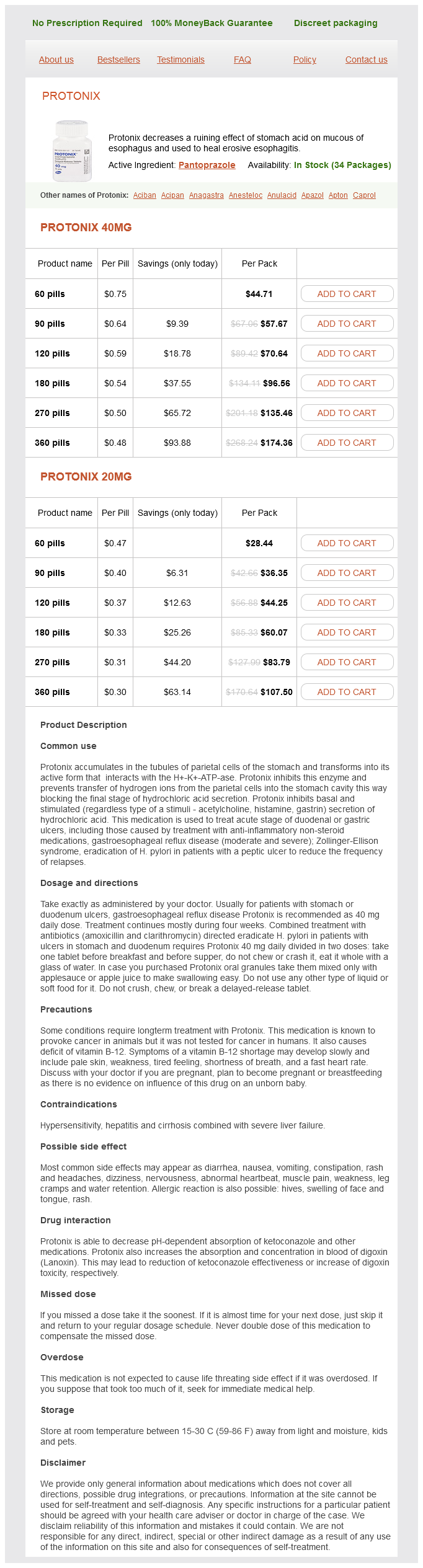
Protonix 40mg
- 60 pills - $44.71
- 90 pills - $57.67
- 120 pills - $70.64
- 180 pills - $96.56
- 270 pills - $135.46
- 360 pills - $174.36
Protonix 20mg
- 60 pills - $28.44
- 90 pills - $36.35
- 120 pills - $44.25
- 180 pills - $60.07
- 270 pills - $83.79
- 360 pills - $107.50
This requires pharmacokinetics and pharmacodynamics studies be conducted in each trimester because physiologic and biologic changes fluctuate throughout gestation gastritis symptoms from alcohol 20 mg pantoprazole visa. Furthermore, these studies need to be performed under various conditions, including singleton and multiple gestations, in fed and fasting states and for long and short durations of treatment. What can be done to overcome the barriers of conducting medication-focused research in pregnancy Although it may seem like an insurmountable problem, we suggest here a few approaches to overcoming the obstacles (described in Current Status of Pharmacotherapy in Pregnancy). First, incentives need to be provided to the manufacturers of these drugs motivating study of medications in pregnancy. This can be accomplished by offering a patent extension for those who comply or by requiring a more diverse population in phases 2 and 3 of drug development studies. Generally, pharmacokinetic, pharmacodynamic and pharmacoepidemiologic studies need to be encouraged in this population. Large exposure registries in pregnancy which facilitate pharmacoepidemiologic research must also be encouraged and financially supported. These large databases can also be used to record rare outcomes and can be accomplished by expanding postmarketing surveillance. Only when these studies are performed more regularly will we have enough data to define the variations of pharmacokinetics and pharmacodynamics in pregnancy. Until this point, when limited pharmacokinetic and pharmacodynamic data exists for a drug to be used in pregnancy, physicians must optimise drug therapy through close monitoring of both the mother and fetus. Medication use during pregnancy, with particular focus on prescription drugs: 19762008. The National Academies Collection: reports funded by National Institutes of Health. Medications in the first trimester of pregnancy: most common exposures and critical gaps in understanding fetal risk. Xenobiotic-induced transcriptional regulation of xenobiotic metabolizing enzymes of the cytochrome P450 superfamily in human extrahepatic tissues. Assessment of the hepatic arterial and portal venous blood flows during pregnancy with Doppler ultrasonography. Body fat and water changes during pregnancy in women with different body weight 16. The pregnancy-related decrease in fasting plasma homocysteine is not explained by folic acid supplementation, hemodilution, or a decrease in albumin in a longitudinal study. Cytochrome P450 enzymes in drug metabolism: regulation of gene expression, enzyme activities, and impact of genetic variation. Pittsburgh: University of Pittsburgh, School of Pharmacy, Department of Pharmaceutical Science; 2015. Pregnancy-induced changes in the pharmacokinetics of caffeine and its metabolites. Prospectively assessed changes in lamotrigineconcentration in women with epilepsy during 29. Prevention of preterm delivery with 17-hydroxyprogesterone caproate: pharmacologicconsiderations. Expression of cytochrome P450 2E1 in normal human bronchial epithelial cells and activation by ethanol in culture. Expression of xenobiotic-metabolizing cytochrome P450 forms in human full-term placenta. Detection of cytochrome P450 gene expression in human placenta in first trimester of pregnancy. Despite improvement in perinatal survival, the rates of shortand long-term morbidity (including neurodevelopmental outcome) remain substantial. Optimising transition in the first minutes after birth by improving lung aeration and applying strategies to delay cord clamping can reduce the risks of lung injury and cerebral injury.
Pantoprazole dosages: 40 mg, 20 mgPantoprazole packs: 60 pills, 90 pills, 120 pills, 180 pills, 270 pills, 360 pills
At the apex gastritis cure home remedies generic 20 mg pantoprazole amex, there was a grade l/6 1 holosystolic murmur, an opening snap, and a grade 2 dia stolic rumble with presystolic accentuation. Chest roentgenogram showed a normal-sized heart, an enlarged left atrium, a mild degree of pulmonary vascular redistribution, and no calcification in the region of the mitral valve, and was otherwise normal. This patient was symptomatic because of her increased left atrial pressure and atrial arrhythmia. She had not yet developed the second stenosis, discussed previ ously, at the precapillary pulmonary arteriolar level. Thus her pulmonary artery pressure elevation was purely a con sequence of the increased left atrial and pulmonary venous pressures, and the pulmonary vascular resistance was normal (< 1 20 dyn-second- cm-5). Without relief of the mitral stenosis, her paroxysmal atrial fibrillation will likely become persistent. She was asymptomatic until she was 19 years old, when dur ing the last month of her first pregnancy, at which time she was quite anemic, she developed pulmonary congestion. She responded well to medical therapy and remained asymptom atic thereafter, even during three subsequent pregnancies. However, during her fifth pregnancy at age 3 7, dyspnea, orthopnea, paroxysmal nocturnal dyspnea, and one episode of hemoptysis of red blood occurred at the seventh month, necessitating hospitalization through term. Thereafter she improved, but became progressively tired with loss of energy and drive. If she pushed herself, she would become somewhat short of breath on a flight of stairs, but it was fa tigue more than breathlessness that bothered her. Her blood pressure was 1 1 5/70 mmHg, and her pulse, 90 beats per minute and irregularly irregular. The neck veins were just visible at the clavicles with the patient sitting upright. The point of maximal impulse was in the fifth interspace just outside the midclavicular line. Chest radiographs showed the heart to be moderately enlarged because of enlargement of the left atrium and right ventricle. The pulmonary arteries were prominent, and there was a mod erate degree of pulmonary vascular redistribution. Her symptoms then improved for nearly 2 years, only to return 2 months prior to admission. Since then, despite a good cardiac regimen, she had had a bed-chair-bathroom existence. Blood pressure was 96/72 mmHg; pulse rate was 90 beats per minute and irregu larly irregular; respirations were 32 per minute. N eck veins were distended to the angle of the j aw, v waves were promi nent, and there were bibasilar rales over the lung fields. An opening snap was present, and there was a barely audible mitral diastolic murmur with appreciable presystolic accentuation. Chest roentgenogram showed a large heart with prominent left atrium, right ventricle, pul monary arteries, pulmonary vasculature, and Kerley B lines. Subsequently, however, her maj or symptom was fatigue, associated with a reduced cardiac output and an increased arteriovenous 0 2 difference. The orthopnea and paroxysmal dyspnea had receded somewhat despite the fact that her pulmonary capillary pressure was at the pulmonary edema level. This is a common, although poorly understood, phenomenon in patients with mitral stenosis when pulmonary vascular disease begins to occur. Thus, this patient was beginning to develop the second ste nosis discussed previously, and this is apparent from the ele vated pulmonary vascular resistance (520 dyn-second- cm-5). As was true for the patient j ust discussed (case 1), appropriate therapy would be either balloon mitral valvuloplasty or surgical valve replacement. At the time of presentation to us, she showed evidence of advanced right heart failure and pulmonary hypertension. This woman has two stenoses, and both are severe: the mitral ori fice area is less than one-tenth the normal at 0. Even at this stage in their course, patients can respond dramatically to correction of their mitral stenosis. Physiology Mitral regurgitation from whatever cause implies a double outlet to the left ventricle: During systole, blood exits the left ventricle through both aortic and mitral valves. Although total left ventricular output rises, forward output into the aorta may fall.
Phosphatidyl Serine (Phosphatidylserine). Pantoprazole.
- Are there any interactions with medications?
- What is Phosphatidylserine?
- Are there safety concerns?
- Depression, exercise-induced stress, improving athletic performance, improving thinking ability, attention deficit-hyperactivity disorder (ADHD), and other conditions.
- What other names is Phosphatidylserine known by?
- Dosing considerations for Phosphatidylserine.
- Confusion in older people (senile dementia).
Source: http://www.rxlist.com/script/main/art.asp?articlekey=96953
Then gastritis symptoms last cheap pantoprazole 20 mg overnight delivery, within another small fraction of a second, the inactivation gates of the channels open, and a new action potential can be initiated. The period during which a second action potential cannot be elicited, even with a strong stimulus, is called the absolute refractory period. Therefore, one can readily calculate that such a fiber can transmit a maximum of about 2500 impulses per second. Inhibition of Excitability-Stabilizers and Local Anesthetics In contrast to the factors that increase nerve excitability, membrane-stabilizing factors can decrease excitability. For example, a high extracellular fluid calcium ion concentration decreases membrane permeability to sodium ions and simultaneously reduces excitability. Among the most important stabilizers are the many substances used clinically as local anesthetics, including procaine and tetracaine. Most of these agents act directly on the activation gates of the sodium channels, making it much more difficult for these gates to open and thereby reducing membrane excitability. When excitability has been reduced so low that the ratio of action potential strength to excitability threshold (called the safety factor) is reduced below 1. The reason for this restriction is that shortly after the action potential is initiated, the sodium channels (or calcium channels, or both) become inactivated, and no amount of excitatory signal applied to these channels at this point will open the inactivation gates. The only condition that will allow them to reopen is for the membrane potential to return to or near the original 40 20 Millivolts 0 20 40 60 80 0 A 1 B C 2 3 Milliseconds D 4 Acute subthreshold potentials Threshold Bibliography Alberts B, Johnson A, Lewis J, et al: Molecular Biology of the Cell, 5th ed. Note the development of acute subthreshold potentials when the stimuli are below the threshold value required for eliciting an action potential. Axo-myelinic neurotransmission: a novel mode of cell signalling in the central nervous system Nat Rev Neurosci. Voltage-gated calcium channels: key players in sensory coding in the retina and the inner ear. In this articler, we mainly consider skeletal muscle function; the specialized functions of smooth muscle are discussed in Chapter 8, and cardiac muscle is discussed in Chapter 9. The light bands contain only actin filaments and are called I bands because they are isotropic to polarized light. The dark bands contain myosin filaments, as well as the ends of the actin filaments, where they overlap the myosin, and are called A bands because they are anisotropic to polarized light. It is the interaction between these cross-bridges and the actin filaments that causes contraction (Video 6-1). From this disk, these filaments extend in both directions to interdigitate with the myosin filaments. The Z disk, which is composed of filamentous proteins different from the actin and myosin filaments, passes crosswise across the myofibril and also crosswise from myofibril to myofibril, attaching the myofibrils to one another all the way across the muscle fiber. Therefore, the entire muscle fiber has light and dark bands, as do the individual myofibrils. The portion of the myofibril (or of the whole muscle fiber) that lies between two successive Z disks is called a sarcomere. At this length, the actin filaments completely overlap the myosin filaments, and the tips of the actin filaments are just beginning to overlap one another. As discussed later, at this length, the muscle is capable of generating its greatest force of contraction. Except for about 2% of the fibers, each fiber is usually innervated by only one nerve ending, located near the middle of the fiber. The sarcolemma consists of a true cell membrane, called the plasma membrane, and an outer coat made up of a thin layer of polysaccharide material that contains numerous thin collagen fibrils. At each end of the muscle fiber, this surface layer of the sarcolemma fuses with a tendon fiber. The tendon fibers, in turn, collect into bundles to form the muscle tendons that then connect the muscles to the bones. Each titin molecule has a molecular weight of about 3 million, which makes it one of the largest protein molecules in the body. Also shown in cross section are T tubules (arrows) that lead to the exterior of the fiber membrane and are important for conducting the electrical signal into the center of the muscle fiber. Part of the titin molecule is closely associated with the myosin thick filament, whereas the rest of the molecule is springy and changes length as the sarcomere contracts and relaxes.
Syndromes
- Slowly return to your regular activities. A physical therapist can help you do this safely.
- Stopping food or fluid by mouth to limit the activity of the pancreas
- Parasites
- Diarrhea
- Nausea
- Joint x-rays
- Opening of the artery with a balloon catheter (angioplasty) with or without a stent
One thing that has been learned gastritis diet ����� order cheap pantoprazole, especially in the setting of car diogenic shock, is that more flow does not necessarily equate with improved survival; clearly, other factors need to be con sidered. It is anticipated that these devices will continue to evolve and, perhaps more importantly, that new studies will address the long-standing question of whether and how these devices can be used most effectively to reduce mortality in cardiogenic shock. Diastolic balloon pumping (with carbon dioxide) in the aorta-a mechanical assistance to the failing circulation. Initial clinical experience with intraaortic balloon pumping in cardiogenic shock. Complications associated with percutaneous placement and use of intraaortic balloon coun terpulsation. Stroke complicating percuta neous coronary interventions: incidence, predictors, and prognos tic implications. Long-term follow-up of patients un dergoing intraaortic balloon counterpulsation and operation. The safety of in traaortic balloon pump catheter insertion through suprainguinal prosthetic vascular bypass grafts. Feasibility and long-term safety of elective lmpella-assisted high-risk percutaneous coronary inter vention: a pilot two-centre study. Transfemoral selective coronary artery catheterization during intra-aortic balloon by-pass pumping. The role of heparin anticoagu lation during intra-aortic balloon counterpulsation in the coronary care unit. Early revasculariza tion in acute myocardial infarction complicated by cardiogenic shock. A randomized clinical trial to evaluate the safety and efficacy of a percutaneous left ventricu lar assist device versus intra-aortic balloon pumping for treatment of cardiogenic shock caused by myocardial infarction. Randomized comparison of intra aortic balloon support with a percutaneous left ventricular assist device in patients with revascularized acute myocardial infarction complicated by cardiogenic shock. A randomized multicenter clinical study to evaluate the safety and efficacy of the TandemHeart percutaneous ventricular assist device versus conven tional therapy with intraaortic balloon pumping for treatment of cardiogenic shock. Meyns B, Stolinski j, Leunens V, Verbeken E, Flameng W Left ven tricular support by catheter-mounted axial flow pump reduces in farct size. Elective intra-aortic balloon counterpulsation during high-risk percu taneous coronary interven tion: a randomized controlled trial. The current prac tice of intra-aortic balloon counterpulsation: results from the Benchmark Registry. Contemporary u tilization and outcomes of intra-aortic balloon counterpulsation in acute myocardial infarction: the benchmark registry. Five-year results of 219 consecu tive patients treated with extracorporeal membrane oxygenation for refractory postoperative cardiogenic shock. Hemodynamic support with a percutaneous left ventricular assist device during stent ing of an unprotected left main coronary artery. Prophylactic versus standby cardiopulmonary support for high risk percutaneous transluminal coronary angioplasty. Emergent applications of car diopulmonary support: a multiinstitutional experience. Emergent percutaneous cardiopulmonary bypass in pa tients having cardiovascular collapse in the cardiac catheterization laboratory. Clinical introduction of the TandemHeart, a percutaneous left ventricular assist device, for circulatory support during high-risk percutaneous coronary intervention. Feasibility study of the use of the TandemHeart percutaneous ven tricular assist device for treatment of cardiogenic shock. Cardiogenic shock complicating acute myocardial in farction: expanding the paradigm. The percu taneous ventricular assist device in severe refractory cardiogenic shock. First experiences with a new miniaturised life support system for mobile percutaneous car diopulmonary bypass. A novel tech nique for intraaortic balloon pump placement via the left axillary artery in patients awaiting cardiac transplantation.
Usage: p.r.n.
Segmental wall motion during one of the preceding sinus beats is compared with that of the postextrasystolic beat and improvement on the potentiated beat as compared with the preceding sinus beat suggests ischemia rather than infarction gastritis what to eat proven 20 mg pantoprazole. It is probably unwise, how ever, to attempt to induce the ventricular extrasystole by manipulating the left ventriculographic catheter during the inj ection of contrast material since such manipulation may cause endocardial staining. O ther types of intervention ventriculography have been used in patients with chronic left ventricular volume over load caused by aortic or mitral regurgitation. In the patient with aortic regurgitation and well-preserved left ventricular function, angiotensin in a dose sufficient to increase left ven tricular systolic pressure by 20 to 50 mmHg causes no change in left ventricular ej ection fraction 21 But if aortic regurgita tion has caused a loss of left ventricular contractile reserve, a similar amount of angiotensin can cause a fall of > 0. Thus, left ventriculography during afterload stress has been used to provide additional information about left ventricular functional capability. Alter natively, intervention ventriculography using sodium nitro prusside has been used in patients with mitral regurgitation, aortic regurgitation, or dilated cardiomyopathy to assess the potential benefit in hemodynamics and ventricular perfor mance during chronic vasodilator therapy. Such extrasystoles can usually be elimi nated or at least minimized by repositioning the catheter. Although short runs of ventricular tachycardia occur dur ing an occasional ventriculogram, they almost always cease promptly when the catheter is removed from the ventricle. Rarely, the ventricular tachycardia caused by ventriculogra phy is sustained even after catheter removal. It should be treated quickly with a bolus of intravenous lidocaine and, if necessary, direct current countershock. Ventricular fibril lation has been reported to be induced by an improperly grounded power inj ector. Although a small endocardial stain usu ally causes no problem, a large stain may lead to medically refractory ventricular tachyarrhythmias, including ventricu lar tachycardia or fibrillation. Rarely, the power inj ection of contrast material causes myocardial perforation, with resul tant leakage of blood and contrast material into the pericar dia! This must be treated by emergency pericardiocentesis and immediate consultation obtained from a cardiothoracic surgeon (see Chapters 3 8 and 44). Complications of Contrast Med ia With earlier ionic contrast agents, ventriculography produced a modest fall in systemic arterial pressure, a reflex increase in heart rate, and a transient depression of left ventricular con tractility that resolved within 1 to 2 minutes. Patients used to experience a hot flash owing to the powerful vasodilation caused by the contrast material as it distributed through out the arterial tree, and nausea or vomiting could occur in 20% to 30% of cases. In the patient with underlying right bundle branch block and left posterior fascicular block, complete heart block may occur as the catheter is advanced into the left ventricle. Transient complete left bundle branch block is an extremely rare complication of retrograde left heart catheterization. In a few subj ects, echocardiography may fail to provide adequate images owing to extreme obesity or an increased anteroposterior chest dimension unless a trans esophageal study is performed, or contrast is used. In most patients, however, adequate images of the left ventricle can be acquired in multiple short- and long-axis planes to evaluate segmental and global left ventricular function as well as the degree of mitral regurgitation. The left ventricular volumes provided by echocardiography tend to be somewhat smaller and the estimates of mitral regurgita tion tend to be somewhat higher than those obtained with contrast ventriculography. N everthe less, the risk of air embolization should be avoidable by fol lowing good practices in filling the inj ector and confirming a bubble-free hookup as described above. The presence of thrombi on or within the ventriculographic catheter is min imized by frequent flushing of the catheter with a solution containing heparin when the ventriculographic catheter is first introduced and just prior to hooking up for the ventricu logram. If there is any suspicion (from noninvasive testing) of a thrombus in the left ventricular apex, great care should be taken to position the ventriculographic catheter in the left ventricular inflow tract, avoiding the apical portion com pletely, or ventriculography itself should be avoided, relying on noninvasive evaluation. Partially organized thrombi may also be dislodged from the left ventricular wall by the catheter tip or the force of a power inj ection. Accordingly, the ven tricular angiographic catheter should not be advanced to the left ventricular apex except under exceptional circumstances. First pass imaging, a technically demanding method based on the transit of a rapid bolus of a radionuclide accompanied by high-temporal resolution image acquisition, may be performed at rest or with exercise and is perhaps the most accurate method for right ventricular functional determination. These images are acquired in a gated fashion throughout the cardiac cycle, and end-diastolic and end-systolic frames are identi fied. Stroke volume, cardiac output, and other hemodynamic parameters may also be obtained. Myocardial tagging overlays a grid onto the myo cardial walls and allows for clear visualization of deformities and wall motion abnormalities, with a warping of the grid pattern indicating how the myocardium twists and stretches as it contracts. Automated determination of myocar dial surfaces in multiple axial images permits assessment of ventricular volume by the method of discs, similar to echo cardiography, but with markedly improved spatial resolution.
References
- Klein F, Amin Kotb WFM, Petersen I. Incidence of human papilloma virus in lung cancer. Lung Cancer 2009;65(1):13-8.
- Lan NC, Karin M, Nguyen T, et al. Mechanisms of glucocorticoid hormone action. J Steroid Biochem 1984;20:77-88.
- Arciniegas DB, Anderson CA. Viral encephalitis: neuropsychiatric and neurobehavioral aspects. Curr Psychiatry Rep. 2004;6(5):372-379.
- Goldstein JL, Brown MS. The LDL pathway in human fibroblasts: a receptor-mediated mechanism for the regulation of cholesterol metabolism. Curr Top Cell Regul 1976;11:147.
- Gori T, Parker JD. Nitrate tolerance: a unifying hypothesis. Circulation 2002;106: 2510-2513.
- Calabrese E. Infrapopliteal arterial diseases: angioplasty and stenting. In: Heuser RR, Henry M, eds. Textbook of peripheral vascular interventions, 2nd ed. London: Informa HealthCare, 2008:633-638.