Ketoconazole Cream
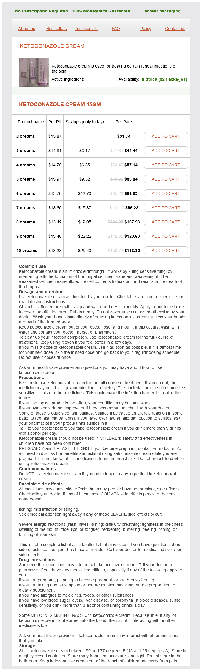
Ketoconazole Cream 15gm
- 2 creams - $31.74
- 3 creams - $44.44
- 4 creams - $57.14
- 5 creams - $69.84
- 6 creams - $82.53
- 7 creams - $95.23
- 8 creams - $107.93
- 9 creams - $120.63
- 10 creams - $133.32
This phenomenon may explain the association between elevated lipoprotein a levels and atherosclerosis antibiotics for strep viridans uti ketoconazole cream 15 gm purchase on line. In contrast, thrombosis reflects a pathologic process associated with intravascular thrombi that fill the lumens of arteries or veins. Hemostatic Disorders Bleeding can occur if there is abnormal platelet plug formation or reduced thrombin generation and subsequent fibrin clot formation at the site of vascular injury; disorders of primary and secondary hemostasis, respectively. Bleeding also can occur if the platelet or fibrin clot is prematurely degraded because of excessive fibrinolysis; a disorder of tertiary hemostasis, the features distinguishing disorders of primary, secondary, and tertiary hemostasis are outlined in Table 124-1. Hemorrhagic disorders can be inherited or acquired, and the clinical and laboratory evaluation of such disorders is detailed in Chapters 130 and 131, respectively. Because the platelet plug provides the first line of defense against bleeding, patients with disorders of primary hemostasis often present with immediate bleeding after injury and petechiae, pinpoint hemorrhages, may be noted. Thrombocytopenia can be the result of decreased production, which can occur because of failure, infiltration, or fibrosis of the bone marrow (see Chapters 27 and 28); increased platelet destruction; or abnormal distribution because of platelet pooling in the spleen (see Chapter 134). Platelet function disorders include disorders of (1) platelet adhesion, such as von Willebrand disease (see Chapter 140) and BernardSoulier syndrome (see Chapter 127); (2) thromboxane synthesis; (3) secretion, such as alpha or dense granule deficiency or aspirin-like secretion defects; (4) aggregation, such as Glanzmann thrombasthenia (see Chapter 127); or (5) procoagulant activity (Scott syndrome) in which the platelets fail to support clotting factor complex assembly (see Chapter 128). Acquired disorders of platelet function can occur in patients taking drugs that impair platelet function, such as aspirin or nonsteroidal antiinflammatory drugs, or in patients with uremia, paraproteins, or myelodysplastic or myeloproliferative disorders (see Chapter 132). Bleeding can also occur with inflammation or malformations of the blood vessels or abnormalities of the connective tissue supporting the blood vessels. Inflammatory disorders include Henoch-Schonlein purpura (see Chapter 152) and the vasculitis that occurs with paraproteins or cryoglobulins or in patients with systemic lupus erythematosus or other immune disorders. Telangiectatic vessels can often be seen in the oral and nasal cavities of patients with this disorder and bleeding episodes, primarily from the nose and gastrointestinal tract, are common. Abnormalities of the connective tissue matrix supporting the blood vessels include Marfan syndrome, Ehlers-Danlos syndrome, and pseudoxanthoma elasticum. Disorders of Secondary Hemostasis Secondary hemostasis depends on rapid generation of sufficient amounts of thrombin to generate a fibrin mesh that not only consolidates the platelet aggregates that form at sites of vascular injury but is also stable enough to provide a barrier that prevents leakage of blood from the damaged blood vessel. The frequency of bleeding episodes in patients with severe hemophilia can be reduced by prophylactic administration of the appropriate factor concentrate; such treatment is also administered to those with hemophilia who have overt bleeding or in preparation for surgery or other major interventions. Management of hemophilia becomes more complicated if patients develop inhibitory antibodies that attenuate or abolish the activity of the infused factor (see Chapter 138). The clinical and laboratory evaluation of such patients are detailed in Chapters 130 and 131, respectively, and their treatment is outlined in Chapter 116. Acquired deficiencies of coagulation factors can result from decreased synthesis because of severe liver disease, vitamin K deficiency or intake of drugs that interfere with vitamin K metabolism, consumption because of excessive activation of coagulation. Congenital disorders of fibrinogen include absence or low levels of fibrinogen (afibrinogenemia and hypofibrinogenemia, respectively) or synthesis of a dysfunctional protein (dysfibrinogenemia). Acquired disorders of fibrinogen include decreased synthesis or production of an abnormal fibrinogen and increased fibrinogen consumption or the presence of inhibitors that interfere with fibrin polymerization, such as paraproteins or autoantibodies, particularly in patients with systemic lupus erythematosus or other immune disorders or elevated levels of fibrin(ogen) degradation products. Stabilization of fibrin requires cross-linking of the and chains of adjacent fibrin monomers to yield a polymer that is resistant to premature breakdown. Incorporation of thrombi into plaques promotes plaque growth, and decreased endothelial cell production of heparan sulfate-which normally limits smoothmuscle proliferation-contributes to plaque expansion. Intracardiac Thrombosis Thrombi can form in the left ventricle after transmural myocardial infarction or with an aneurysm or dyskinetic ventricle or in the left atrial appendage, particularly in patients with atrial fibrillation (see Chapter 149). Damage to the endothelium after myocardial infarction and abnormal blood flow are the major triggers for left ventricular thrombus formation. With rapid atrial fibrillation, there also is stasis and turbulent blood flow in the left atrial appendage, which is a long, blind-ended trabeculated pouch. The generation of thrombin creates a local hypercoagulable state that likely promotes thrombus formation on the abnormal endothelium. Embolization of these thrombi to the brain is a common cause of ischemic stroke and is the major cause of mortality and morbidity in patients with atrial fibrillation. Disorders of Tertiary Hemostasis Tertiary hemostasis depends on the generation of plasmin, which degrades fibrin and restores blood flow in damaged vessels. Premature lysis of fibrin in hemostatic plugs can lead to bleeding; this can occur systemically or can be localized (Table 124-4). More commonly, systemic hyperfibrinolysis is secondary to activation of coagulation by procoagulants such as tissue factor.
Ketoconazole Cream dosages: 15 gmKetoconazole Cream packs: 2 creams, 3 creams, 4 creams, 5 creams, 6 creams, 7 creams, 8 creams, 9 creams, 10 creams
Immediate Thrombocytopenia After Liver Transplantation More than 90% of adults have been reported to have thrombocytopenia within the first week after liver transplantation antibiotic resistant bacteria cure order ketoconazole cream online from canada. An increase in thrombopoietin is seen on days 4 to 6, and increased reticulated platelets are noted on days 7 and 8. Lack of resolution is associated with a poor prognosis for graft and overall survival. One theory is that it is a nonimmune consumptive process reflected by increased markers of thrombin generation. A second theory is that it is unlikely to be a consumptive process because of the lack of platelet activation after day 1. The thrombocytopenia is more likely a result of low levels of thrombopoietin seen pretransplant. With new production and rising levels of thrombopoietin from the new liver, there is a subsequent increase in platelet production, resulting in normalization of the platelet count soon after transplantation. Although the specific etiology has not been clarified, there is general consensus that it is not immune mediated. Although originally azathioprine was believed to be a cause of this thrombocytopenia, there is no evidence that alteration of azathioprine dose is necessary. Delayed Posttransplantation Thrombocytopenia Delayed thrombocytopenia is not a major problem after solid organ transplantation, and most of the data are published in adult series. In one retrospective study of 36 adult liver transplant recipients, mild thrombocytopenia (<140,000/µL) was seen in 54% of patients at 1 year and 25% at 3 years after transplant. In one report, three of 25 (12%) pediatric liver transplant recipients were found to have isolated thrombocytopenia. In an additional study of 126 children who underwent cardiothoracic transplantation, nine (8%) were noted to have isolated thrombocytopenia. Sirolimus has been associated with mild thrombocytopenia in adults, although the reported frequency has varied. In a study of renal transplant recipients, 23% were reported to have mild thrombocytopenia,293 but no liver transplant patients taking the drug were found to be thrombocytopenic. Steroids provided effective therapy in four and rituximab in four, although splenectomy was ultimately necessary in three. Acquired Glanzmann thromboasthenia has been reported in two children after cardiac transplantation. One patient had multiple autoantibodies, and the other subsequently developed additional antiplatelet antibodies. Alloimmune thrombocytopenia was reported in three recipients of organs from the same donor (two kidneys and a liver). Treatment of Immune-Mediated Thrombocytopenia After Solid Organ Transplantation in Children There is no standard approach to the treatment of immunemediated thrombocytopenia that occurs after solid organ transplantation. First, all other causes of thrombocytopenia should be ruled out before beginning therapy for what is often a presumptive diagnosis of immune-mediated thrombocytopenia. If the thrombocytopenia is mild to moderate (>20,000-30,000/µL) with no associated bleeding symptoms, we usually observe without intervention and monitor the platelet count on at least a weekly basis. If the platelet count is lower than 10,000 to 15,000/µL or if there is bleeding, immediate treatment is initiated with high-dose corticosteroids: 4 mg/kg/day of prednisone (or intravenous equivalent) divided in three or four doses and continued for 4 days. For patients who have recurrent or chronic thrombocytopenia requiring multiple courses of treatment to maintain platelet counts greater than 10,000 to 15,000/µL, we have used vincristine (1. Involvement of both the transplant team and the hematology team is necessary for ideal management of these complex patients. Immune-Mediated Thrombocytopenia Posttransplantation There are many case reports of immune-mediated thrombocytopenia after all types of solid organ transplantation in children and adults and either alone or in combination with other cytopenias. Eight Chapter 154 Hematologic Manifestations of Childhood Illness 2161 Infection-Associated Thrombocytopenia Posttransplantation Although there are few published reports, one would expect the same hematologic problems, including thrombocytopenia, associated with bacterial sepsis or other serious infections as seen in nontransplant patients. These and other symptoms of herpesvirus infection occur at the same frequency as nontransplant patients. There is a case report of a child after liver transplant who developed measles complicated by autoimmune thrombocytopenia and neutropenia. Filgrastim has been effective in reversing the neutropenia, although some patients required decreasing the dose or stopping the drug.
MCP (Pectin). Ketoconazole Cream.
- How does Pectin work?
- What other names is Pectin known by?
- Diarrhea, reducing the risk of colon cancer, diabetes, infection, mouth and throat sores, reducing damage from radiation, preventing heavy metal toxicity, prostate cancer, heartburn, and other conditions.
- Are there any interactions with medications?
- High cholesterol.
- Are there safety concerns?
Source: http://www.rxlist.com/script/main/art.asp?articlekey=96506
This lymph node demonstrates large non-cohesive large cells with extensive necrosis antibiotics for uti how long buy ketoconazole cream 15 gm mastercard. These processes form the fundamental mechanisms by which cell function, cellcell interactions and cell turnover are regulated. Disruption of this regulation may lead to disease, whilst an understanding of these mechanisms allows the physician to attempt to predict disease behaviour and to explore methods of restoring this regulation at a molecular level. This chapter reviews the principles of molecular genetics and outlines aspects of the molecular biology of the cell in the context of otolaryngological disease processes and describes some of the techniques that form the backbone of current molecular biology. It should give the reader sufficient background knowledge of molecular biology to understand subsequent chapters discussing the molecular biology of specific otolaryngological conditions. The nucleotides on each strand are organized linearly in triplets, known as codons. However, as there are more triplet combinations (64) than commonly encountered amino acids (20), some proteins may be represented by different codons. The mitochondrial genome and its potential role in cancer diagnostics will be discussed later. One of each pair of chromosomes is maternally inherited and the other is paternally inherited. Each chromosome has a distinctive shape, size and banding pattern, but have the common appearance of two arms apparently separated by a constriction. The centromere is microscopically recognizable as the central constriction separating the chromosome into a long arm (q for queue) and a short arm (p for petit), but its biological role lies in anchoring the chromosome to the mitotic spindle for segregation during cell division. The ends of the chromosomes are capped by telomeres, which are specialized structures containing unique simple repetitive sequences. They maintain the structural integrity of the chromosome and provide a solution for complete replication of the extreme ends of the chromosome. The conventional nomenclature for chromosomal locus assignment is given by the chromosome number, followed by the arm and finally the position on the arm, for example, 3p21 indicates position 21(two-one) on the short arm of chromosome three. The specificity of the complementary relationship between the nucleotides on each strand forms the basis for many techniques of modern molecular biology and molecular cytogenetics. The exact function of these microsatellite repeats is not entirely clear, but their existence and frequency of dispersion throughout the genome have greatly facilitated study of the genetics of tumours and many inherited disorders, which will be discussed later. The methylation status of these CpG islands determines whether that gene is expressed in a particular cell or tissue, being unmethylated in tissues where the genes are expressed. As will be discussed later, aberration of this control is one of the mechanisms of tumour suppressor gene inactivation. The polypeptide subsequently undergoes a variable degree of post-translational modification and/or cleavage to produce the mature protein product, which may have an intracellular role or may be exported to the endoplasmic reticulum and hence to the extracellular space to execute its function. Unlike the nuclear Chapter 1 Molecular biology]5 genome, which is inherited from mother and father, the mitochondrial genome of an individual is entirely maternally inherited. By surveying many different bacteria, a wide range of restriction endonucleases is now available, each of which recognize specific target sites based on sequences of four to eight nucleotides. As a specific, seven nucleotide sequence (heptanucleotide) will occur less frequently than a four nucleotide sequence (tetranucleotide), statistically, endonucleases recognizing heptanucleotide targets will cut less frequently thereby yielding larger fragments than those recognizing tetranucleotides. Chromosome locus nomenclature: chromosome number 3p21 position on chromosome arm. This allows fragments of different sizes to be resolved within a suitable gel matrix by the application of an electric current across the matrix. The choice of the particular matrix depends on the fragment sizes that one is trying to resolve. Polyacrylamide gels can resolve differences of just one base pair between fragments of several hundred base pairs in size by virtue of a small pore size in the gel matrix. Beyond that size, electrophoretic mobility is no longer proportional to fragment size. This elegantly simple process is eponymously known as Southern blotting after the scientist who described the process in 1975. Two other commonly used transfer techniques have their names derived from Southern blotting as jargon terms. Western blotting is one of the mainstays of protein analysis and involves the transfer of electrophoresed protein bands from a polyacrylamide gel on to a nitrocellulose or nylon membrane to which they bind strongly.
Syndromes
- Bronchoscopy -- camera down the throat to see burns in the airways and lungs
- Blurred vision
- Pelvic CT scan
- Shortness of breath
- CT scan of the abdomen and pelvis
- It is common for children to spend up to a week in the hospital. Sometimes a cast is placed on the leg for 3 to 4 weeks.
- The pain may feel like a deep ache, or a shooting, burning pain.
- Mental deterioration
- Poor healing of the wounds
- 0.10 -- slurred speech
Diaz-Ricart M antibiotics for acne for 6 months purchase ketoconazole cream master card, Etebanell E, Cases A, et al: Erythropoietin improves signaling through tyrosine phosphorylation in platelets from uremic patients. Violi F, Ferro D, Basili S, et al: Prognostic value of clotting and fibrinolytic systems in a follow-up of 165 liver cirrhotic patients. Violi F, Ferro D, Basili S, et al: Hyperfibrinolysis increases the risk of gastrointestinal hemorrhage in patients with advanced cirrhosis. Violi F, Leo R, Basili S, et al: Association between prolonged bleeding time and gastrointestinal hemorrhage in 102 patients with liver cirrhosis: Results of a retrospective study. Laffi G, Cominelli F, Ruggiero M, et al: Altered platelet function in cirrhosis of the liver: Impairment of inositol lipid and arachidonic acid metabolism in response to agonists. Kunihiro N, Kawai B, Sanjo A, et al: Platelet aggregation and coagulation and fibrinolysis parameters in both portal and systemic circulations in patients with cirrhosis and hepatocellular carcinoma. Tripodi A, Primignani M, Chantarangkul V, et al: An imbalance of pro- vs anti-coagulation factors in plasma from patients with cirrhosis. Cortelazzo S, Viero P, Casarotto C, et al: Bleeding on patients with autoimmune thrombocytopenic purpura and normal platelet count. Clancy R, Jenkins E, Firkin B: Qualitative platelet abnormalities in idiopathic thrombocytopenic purpura. Berchtold P, Muller D, Beardsley D, et al: International study to compare antigen-specific methods used for the measurement of antiplatelet autoantibodies. Takahashi R, Sekine N, Nakatake T: Influence of monoclonal antiplatelet glycoprotein antibodies on in vitro human megakaryocyte colony formation and proplatelet formation. Sugiyama T, Okuma M, Ushikubi F, et al: A novel platelet aggregating factor found in a patient with defective collagen-induced platelet aggregation and autoimmune thrombocytopenia. Deckmyn H, Van Houtte E, Vermylen J: Disturbed platelet aggregation to collagen associated with an antibody against an 85- to 90-Kd platelet glycoprotein in a patient with prolonged bleeding time. Toltl, Ishac Nazi, James Smith, and John Kelton Platelets are anucleate cells that are required for primary hemostasis. This primitive feedback system is very effective at maintaining the platelet count at a stable level. Immune-mediated platelet disorders disrupt normal regulation of platelet number because of cell-mediated or antibody-mediated platelet destruction or megakaryocyte injury. Antibodies that target self (autoimmune) or nonself (alloimmune) antigens on platelets can cause severe thrombocytopenia. These platelet disorders have related features yet are distinct clinical syndromes (Table 133-1). The pathophysiology, clinical manifestations, and management of each of these disorders are discussed in this chapter. Seasonal variability suggests that a viral infection may trigger the disease in many children. Most studies in children report an overall male predominance in early childhood and equalization or reversal to female predominance in older children. Relatively stable incidence rates were found in women up to the age of 60 years with a steady increase thereafter. The prevalence in women is reported to be nearly double that in men; however, this trend is attenuated in older age groups and may revert to a male predominance after the age of 65 years. Adult incidence and prevalence rates may reflect a true increase in risk of disease with age or measurement bias because of a higher likelihood of discovering incidental thrombocytopenia with more frequent medical visits. Recently, however, it has become evident that relative platelet underproduction is also an important feature of this disorder. It is characterized by mild thrombocytopenia with platelet counts generally greater than 70 × 109/L, occurs late in pregnancy, and is clinically insignificant for the mother or baby. Pregnancy-related vascular disorders are the next most common cause of thrombocytopenia during pregnancy. Platelet counts tend to be mildly reduced, and hypertension and other features are invariably present. Immune thrombocytopenia is an uncommon cause of thrombocytopenia in pregnancy, and when mild, it can be difficult to distinguish from incidental thrombocytopenia of pregnancy. Epidural anesthesia is not recommended unless the platelet count is above 70 to 80 × 109/L, although this practice is operator driven and based on little evidence.
Usage: ut dict.
It is important to realize that the coagulation proteins need to decrease to different levels before the various screening assays show an abnormality antibiotics for uti uti cheap 15 gm ketoconazole cream overnight delivery. The sensitivity of the screening tests for detection of specific abnormalities varies with the factor being tested, the commercial reagent used in the assay, and the equipment platform for measurement. In the coagulation cascade hypothesis, coagulation proteins are classified as members of the intrinsic system, the extrinsic system, or the common pathway. In these assays, a sequence of proteolytic reactions takes place, leading to thrombin formation and its proteolysis of fibrinogen. Proteolysis of fibrinogen results in clot formation with precipitation of soluble proteins that are detected by either increased impedance or turbidity or decreased optical clarity, based on the instrumentation used to measure the result. Furthermore, because a series of reactions must occur to result in the final clot formation, any substance. Knowing what each test measures, the clinician can use the fol lowing approach to evaluate bleeding risk in patients who have pro longed values in one or more of these assays (Table 1311). It also is important to recognize these protein defects so that patients do not undergo unnecessary plasma replacement therapy. Individuals can have acquired disorders that inhibit specific coagu lation factors and increase their risk for bleeding. The clinical labora tory phenotype of these patients depends on the protein to which the coagulation protein inhibitor is directed. Inhibitor titer is determined by the degree of dilution of test plasma to obtain a factor level of 50% normal. A complete discussion on performance and interpretation of this assay is seen in Schmaier and Miller. Anticoagulation, unless the patient is faking medical illness, and massive transfusion are excluded by history. The reptilase time uses a snake venom enzyme to "clot" fibrinogen by liberating only fibrinopeptide A. Acquired deficiencies or inhibitors are also seen in a number of medical conditions. Hypergammaglobulinemic states seen with multi ple myeloma or Waldenström macroglobulinemia (immunoglobulin M) can be associated with inhibitors to coagulation protein func tion. This coagulation test pattern is characteristic of a specific inhibitor to a coagulation protein. Another acquired inhibitor that influences coagulation protein reactions is the lupus anticoagulant (see Chapter 143). This inhibitor represents antibodies directed to epitopes of proteins bound to certain phospholipids. Detection of the degree of interference depends on the nature of the commercial reagent. Characteristically a lupus anti coagulant will have a greater effect on a coagulation assay as the reagents, not protein, in the assay are diluted out. After incubation for more than 5 minutes, the time to clot is initiated by recalcification with calcium chloride. After incubation for more than 5 minutes, the time to clot is initiated by addition of calcium chloride. They are simpler to perform than antigen assays for each of the coagulation proteins. The most useful means for determining the presence of an abnormal fibrinogen (dysfibrinogenemia) is to measure clottable fibrinogen and fibrinogen antigen on the same sample. In general, chromogenic assays precisely measure the activity of the protein of interest. The drawback of coagulantbased assays is that the levels or function of other proteins influence the results. For example, a heterozygous factor V Leiden polymorphism gives an abnormal protein C coagu lant assay but will have no influence on protein C activity if per formed by chromogenic assay.
References
- Higgins CB, French JW, Silverman JF, et al: Interruption of the aortic arch: Preoperative and postoperative clinical, hemodynamic and angiographic features. Am J Cardiol 1977; 39:563-571.
- Mehta D, Malik AB: Signaling mechanisms regulating endothelial permeability, Physiol Rev 86:279-367, 2006.
- Kori SH, Foley KM, Posner JB. Brachial plexus lesions in patients with cancer: 100 cases. Neurology 1981; 31(1):45-50.
- Stinchcombe TE, Morris DE, Lee CB, et al. Induction chemotherapy with carboplatin, irinotecan, and paclitaxel followed by high dose three-dimension conformal thoracic radiotherapy (74 Gy) with concurrent carboplatin, paclitaxel, and gefitinib in unresectable stage IIIA and stage IIIB non-small cell lung cancer. J Thorac Oncol 2008;3(3):250-257.
- Kaefer M, Pabby A, Kelly M, et al: Improved bladder function after prophylactic treatment of the high risk neurogenic bladder in newborns with myelomeningocele, J Urol 162(3):1068-1071, 1999.
- Bono AV, Benvenuti C, Gibba A, et al: Adjuvant chemotherapy in locally advanced bladder cancer. Final analysis of a controlled multicentre study, Acta Urol Ital 11(1):5n8, 1997.
- Chawla LS, Davison DL, Brasha-Mitchell E, et al. Development and standardization of a furosemide stress test to predict the severity of acute kidney injury. Crit Care. 2013;17(5):R207.
- Hossmann KA. Pathophysiology and therapy of experimental stroke. Cell Mol Neurobiol 2006;26:1057-83.