Etoricoxib
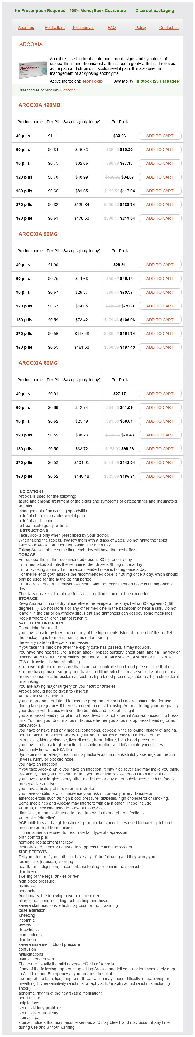
Arcoxia 120mg
- 30 pills - $33.26
- 60 pills - $50.20
- 90 pills - $67.13
- 120 pills - $84.07
- 180 pills - $117.94
- 270 pills - $168.74
- 360 pills - $219.54
Arcoxia 90mg
- 30 pills - $29.91
- 60 pills - $45.14
- 90 pills - $60.37
- 120 pills - $75.60
- 180 pills - $106.06
- 270 pills - $151.74
- 360 pills - $197.43
Arcoxia 60mg
- 30 pills - $27.17
- 60 pills - $41.59
- 90 pills - $56.01
- 120 pills - $70.43
- 180 pills - $99.28
- 270 pills - $142.54
- 360 pills - $185.81
The pharmacokinetic profiles of dexamethasone and methylprednisolone concentration in perilymph and plasma following systemic and local administration arthritis pain and relief buy etoricoxib us. Induction of tolerance by oral administration of beta-tubulin in an animal model of autoimmune inner ear disease. Dobie Noise and aging are responsible for most cases of permanent hearing loss in the United States. The otolaryngologist is expected to verify the existence and severity of the hearing loss, to render a differential diagnosis, to recommend treatment and rehabilitation, to counsel the worker, and to report to referring parties. The goal of this chapter is to provide the otolaryngologist with most of the information needed to meet these needs. Noise-exposed mice may show hearing loss that progresses more rapidly than expected after the noise stops, but this phenomenon has only been demonsttated for a single inbred strain exposed as juveniles (6). Unfortunately, there was no documentation that the men with audiomettic notches had actually been noise exposed (or that those without notches had not). A subsequent longitudinal study &om South Carolina showed no difference in age-related progression of hearing loss between retirees who had had noisy careers and those who had had quiet careers (8), in agreement with the expert consensus. We have already mentioned several explanations for the familiar 4-kHz notch (which can also be at 3 or 6kHz): the greater sensitivity of the human ear to frequencies between 1 and 5 kHz, the protective effect of the acoustic retia: below 2 kHz, and the fact that intermittency is most protective for low frequencies. Recently, another reason has been added: Outer hair cells at the base of the cochlea are especially susceptible to oxidative stress 12). Clearly, inner ear injury and hearing loss are not simply related to the total amount of sound enetgy entering the ear. As many other studies have shown, when workers are exposed to typical broad-spectrum industtial noise, the earliest changes are in the high frequencies (3 to 6kHz). After about 10 years, loss in the high frequencies tends to plateau, but the loss continues to broaden gradually into lower frequencies. Ste reocilia) loss of ganglion cells, may also occur, probably caused by glutamate excitoto:xicity (17). It is generally believed that in these instances sound mechanically damages the organ of Corti. At extreme levels, such as with explosive or blast injury, even tympanic membrane or ossicular injury can occur, causing conductive or mixed hearing loss. The levels at which continuous noise and tones (in contrast to impulses and impacts) cause acoustic trauma are not well defined. The earliest cordless telephones rang through the earpiece; if the person answering the phone failed to switch manually from. Dozens of cases of permanent hearing loss occurred from singlering exposures, each probably less than 1 second in duration. Although almost all cell types may be affected, the outer hair cells are most susceptible to damage. These floppy ste:reodlia may recover their nonnal mechanical properties and function nonnally again. The prinwy site of injury appecu:s to be the rootlets that connect the stereocilia with the top of the hair cell (16). When this question has been addressed explicitly, the bulk of evidence favors simple additivity. Recent federal audiometric swveys, like those in earlier decades, show that age-related hearing loss disproportionately affects higher frequencies, especially in merli varies markedly across individuals; and accelerates in middle age (23,24). Drugs and Chemicals Humes (26) reviewed many studies and concluded that no substantial risk of hearing loss occurs from combining a noise exposure and an aminoglycoside drug when neither is present in sufficient amount to cause hearing loss on its own. Naturally, one would be reluctant to combine ototoxic drugs (primarily aminoglycosides and platinumbased antineoplastia) with hazardous noise exposures, but. The ototoxic drug most likely to be combined with industrial noise exposure is aspirin. Workers exposed to toluene or xylene in addition to noise developed more hearing loss than those exposed to noise alone (27,28). One study (29) showed that styrene exposure below currently recommended values, plus noise, was associated with more hearing loss than noise exposure alone. Men often display more hearing loss in noisy occupations than do women, but this may be due to different nonoccupational exposures (especially shooting) between the genders. Median threshold shifts are shown; half of all workers would show more shift, and half would show less shift than indicated. Hearing threshold levels at age 70 (65-74 years) in the unscreened older adult population of the United States, 1959-1962 and 1999-2006.
Etoricoxib dosages: 120 mg, 90 mg, 60 mgEtoricoxib packs: 30 pills, 60 pills, 90 pills, 120 pills, 180 pills, 270 pills, 360 pills
While sidewall tensioning is worthwhile in virtually any case of lower sidewall laxity arthritis in fingers australia etoricoxib 90 mg without a prescription, the need for structural augmentation grafts cannot always be eliminated with tensioning alone, and an individualized and graduated approach to sidewall reconstruction is still required. In severe cases, the goal of avoiding excessive bulk or airway encroachment and simultaneously achieving adequate sidewall tone and contour is best achieved by aggressive tensioning of the nasal sidewall followed by the judicious and step-wise application of thin, yet rigid augmentation grafts-but only when tensioning fails to provide adequate structural support. Enhancing structural support while limiting graft volume not only optimizes airway patency, it also facilitates a more delicate and refined nasal tip contour, which better suits the aesthetic preferences of the typical revision rhinoplasty patient. One the most common problems encountered in revision of the overresected nasal tip is cephalic retraction of the alar rim following an overzealous cephalic trim procedure. The severity of iatrogenic alar retraction varies widely, ranging from mild to severe. Often these lesser deformities will respond favorably to a combination of sidewall tensioning and alar rim grafts, thereby obviating the need for traditional structural grafts. B,C: Lateral and basal views of alar rim grafts after suture flxatfon to tip complex. E Chapter 184: Revision Rhinoplasty 3021 stability against lobular pinching and/or mild alar retraction (59,69,70). Alar rim grafts typically extend from the nasal dome medially to the mid ala laterally, and can be fashioned from septal, concha], or costal cartilage donor material. Edges should be beveled to optimize graft camouflage and grafts wider than 3 to 4 mm are seldom necessary. Owing to the diminutive size and (nonanatomic) placement away from the internal nasal valve, airway impingement is avoided and external valve rigidity is greatly increased. Treatment of mild alar retraction with alar rim grafts is enhanced by concomitant sidewall tensioning, which serves to tighten and straighten the lateral crural remnant and to oppose upward displacement of the alar rim. Although alar rim grafts are often necessary in the previously operated nose, the small graft size reduces the demand for large amounts of donor cartilage. And by using nasal sidewall tensioning to initially stabilize and tighten the lateral crural remnants, coupled with alar rim grafts to reinforce the nostril rim against cephalic scar contracture, a large number of complex: tip revisions can be effectively managed without any need for ex:tranasal graft material. In addition to avoiding the time, expense, and potential side effects associated with the harvest and placement of extranasal structural grafts, septal donor cartilage offers the additional advantage of a structurally sound, yet characteristically delicate and slender tip contour, better suited to a narrow and refined nasal aesthetic (see Case Four). Although sidewall tensioning is easiest when the (tensioned) crural length coincides exactly with optimal tip projection, sidewall tensioning can be accomplished in virtually any nose by first altering crural length to accommodate overprojected or underprojected alar cartilages. Alterations in crural length can be achieved through a host of conventional rhinoplasty techniques including the lateral crural steal technique for the underprojected tip (71), the lateral crural overlay or vertical lobule division technique for the overprojected tip (72, 73), and the tongue-in-groove technique for both the over- and the underprojected tip (66). In conjunction with tensioning of the crural remnants, conventional suture techniques such as dome sutures, crural spanning sutures, or crural flattening sutures can also be used to further refine alar cartilage contour, while simultaneously preserving crural volume and further reducing dependence upon large structural grafts. Unlike the cephalic trim approach, these tissue-sparing techniques are not predicated upon further narrowing of the lateral crus, and the residual structural integrity of the alar cartilage is fully preserved. Finally, a variety of nonstructural cartilage grafts are also commonly needed to optimize nasal tip contour, symmetry, and alignment in the previously operated nose. A discussion of the myriad strategies used for tip grafting used in cosmetic rhinoplasty is far beyond the scope of this chapter, but primary rhinoplasty grafting techniques are equally effective in revision nasal surgery. Perhaps the most devastating dorsal deformity prompting revision rhinoplasty is overresection of the nasal dorsum, a preventable technical error that may profoundly disrupt nasal bridge aesthetics. Depending upon both the extent and location of overresection, unsightly contour deformities may involve the bony upper vault, the cartilaginous middle vault, or the entire nasal bridge. Unlike the rigid bony dorsum that tends to resist deformation from soft tissue scarring, the comparatively soft and pliable cartilages of the middle vault are particularly susceptible to pinching and collapse from progressive scar contracture. Even when dorsal hump reduction avoids profile overresection, loss of (dorsal) septal width can lead to curvature or deviation of the dorsal septum, collapse or pinching of one or both upper lateral cartilages, or combinations therein. Proactive measures to augment and stabilize middle vault architecture are generally effective in avoiding such problems, but are often neglected at the time of primary rhinoplasty. Moreover, significant functional disturbances are also a common manifestation of nasal bridge deformities. Although the middle vault is considerably more susceptible to postsurgical scar contracture, overaggressive reduction of the bony nasal vault may also lead to conspicuous cosmetic deformities that are especially difficult to treat 3022 Section X: Facial Plastic and Reconstn. In addition to an underprojected and scooped bony profile, overaggressive reduction of the bony pyramid also leads to a widened and washed-out bony dorsum on frontal view. Variations of this characteristic appearance develop when fault¥ lateral osteotomies result in collapse, deviation, and/or comminution of one or both nasal bones.
Ivy-Leafed Cyclamen (Cyclamen). Etoricoxib.
- How does Cyclamen work?
- Menstrual complaints, "nervous emotional states," and digestive problems.
- Dosing considerations for Cyclamen.
- Are there safety concerns?
- What is Cyclamen?
Source: http://www.rxlist.com/script/main/art.asp?articlekey=96431
The interruption of the orbicularis oculi can occasionally contribute to corneal exposure problems exercises for arthritis in back and neck 90 mg etoricoxib order with visa, which usually resolve as the muscle regains functionality. In the interim, corneal protection can include the use of eye lubricants and gentle massage. The presence of corneal exposure from lagophthalmos can be the result of over aggressive skin excision, which is especially problematic in revision upper blepharoplasty or in patients with known dry eye syndrome. In extreme cases, this can be treated with skin grafting, but at the expense of an ideal cosmetic result. Blindness following upper lid blepharoplasty is extremely rare and can result from postoperative hematoma formation. If recognized in a timely fashion, it may be possible to evacuate the hematoma and restore vision. In managing and diagnosing complications, it is important that the patient have easy access to medical care and examination. The patient should be counseled about what is normal after surgery and what is not normal. It is expected that the patient will experience tenderness at the surgical site and some blurry vision from any ointment used. Abnormal postoperative events would be for the eye to swell shut, for pain to be severe, or for there to be any true vision change or loss. It is critical to properly diagnose any underlying brow descent or ptosis prior to upper blepharoplasty in order to offer patients the correct surgical treatment for their complaint. Taking a careful preoperative history and performing an attentive physical examination is important to ensure good outcomes. Careful diagnosis and attention to detail will help avoid any untoward complications and minimize patient dissatisfaction. Strategies for a successful rorrective Asian blepharoplasty after previously failed revisions. Should formal ophthalmologic evaluation be a preoperative requirement prior to blepharoplasty Perkins Jess Prischmann Lower eyelid blepharoplasty is one of the most common and technically challenging procedures in facial plastic surgery. Unlike upper eyelid blepharoplasty, surgery on the lower eyelid has a lower threshold for error and a higher potential for complications. While the skin of the eyelid is the thinnest in the body, the thickness of the orbicularis is variable. The capsulopalpebral fascia is part of the lower eyelid retractor and is an extension of the inferior rectus muscle. The lower eyelid is critical in creating a natural ·almond· shape to the palpebral fissure. It is most commonly angled slightly upward, with the lateral canthus being 2 mm higher than the medial canthus. The central and lateral fat pads are separated by the arcuate expansion of Lockwood ligament the medial fat pad is most commonly transposed to efface the "tear trough deformity" (as described below). The inferior oblique muscle can be vulnerable to injury during lower lid blepharoplasty. It takes a posterolateral course, passes inferior to the inferior rectus muscle, and inserts on the globe. Injury to the inferior oblique can result in a range of disability, from transient diplopia to permanent strabismus. This complication can largely be avoided by only transecting fat that protrudes anterior to the orbital septum (1). The blood supply to the lower eyelid is through rich anastomoses between the internal and external carotid artery systems. The major arteries include the dorsal nasal, angulru; infraorbital, transverse facial, zygomaticofacial, and medial palpebral. From a surgical perspective, the plane between the orbital septum and orbicularis is relatively avascular.
Syndromes
- Nitrocine
- Depression
- Deeply lined face
- Stunted growth in height (short stature)
- An embedded intrauterine device (IUD)
- Tube through the mouth or nose into the stomach to wash out the stomach (gastric lavage)
- Your doctor or nurse will tell you when to arrive at the hospital.
- Abnormal bleeding is accompanied by other symptoms, such as pain, fatigue, dizziness
- Bulging fontanelles in babies
- Brain infection like meningitis or encephalitis, or abscess
There is no need for neutralization of the peeling solution as the frost represents a completed reaction arthritis in fingers natural treatment cheap etoricoxib 120 mg buy online, which is precipitation of 1he keratin by the phenol. Any tearing should be immediately dried to avoid the tea11 pooling the peeling solution into the eye. If not adequately anesthetized, the patient will experience an immediate burning sensation for 15 to 20 seconds. The longer-lasting effect of 1he bupivacaine will greatly aid in minimizing the burning sensation in the postoperative period. In 1he case of excess phenol exposure to the patient or the staff, propylene glycol, glycerol, olive oil, castor oil, or cottonseed oil should be poured onto the site to solubilize the phenol. If exposure to the ey1:8 oa::ua, mineral oil should be immediately applied to the eyes using an eye dropper. When the frost subsides and only erythema persists, a thick layer of bland emollient should be applied to all peeled areas, leaving no peeled skin exposed. None of these topical agents act as an occlusive dressing, and they will therefore not increase the depth of penetration. Starting with postoperative day 1, the patient will apply 1he cream three to four times a day to those areas that are exposed. By using the emollient, the peeled skin can be monitored with greater ease on a dayto-day basis. The patient should have been preoperatively prepared for the expected postoperative edema, erythema, and eventual desquamation. Additionally, patients should also be advised of the burning sensation that may last up to 8 hours following the procedure. Next, the epidermis will begin to change in appearance, becoming leathery, and will separate from the dermis. This reepithelialization will be represented by a conversion of bright red e:rythema to a lighter shade of pink. This final stage of fibroplasia begins within the first week and continues for 12 to 16 weeks after the peel. Strict avoidance of direct, prolonged sun exposure should be encouraged for that 12-week period. It has been the authors experience that sunscreens should also be avoided for the first 6 weeks. Care mun be taken to peel this area so as to not leave a discrete line of unpeeled skin. A comprehensive understanding of these complications and their management will senre the peeler well. Though no death due to phenol peeling has been described in the literature, the possibility of a potential cardi<m. Scular crisis has led to great fear regarding the use of phenol Even in patients preoperatively screened and well hydrated, a reversible arrhythmia can o~ especially in patients with undiagnosed myocardial sensitivity. These patients will develop a supraventricular tach:youdia within 30 minutes of the onset of the peel, which if exacerbated. Increased circulating estrogena can result in hyperpigmentation following chemexfoliation. Dramatic Improvement In rhytlds can be appredated In this patiQnt who undQtoWnt a full. AnoUter example of typical expec:ted results in rhytids and dyschromia after a full·face Hatter chemical peel. Another example of Ute use of a lower acton oil concantration Hetter peel fonnulllion to improve facial dyschromia. In the rare cases in which the cardiac rhythm does not return to normal, proper measures should be employed to treat the aberrant rhythm. Although not as concerning as arrhythmias, prolonged healing times is a nuisance both for the patient and the practitioner. These areas should be treated promptly or they will result in increased risk of scarring. In the event that scarring occurs, it is going to occur most often in the perioral area, specifically the upper lip. Scarring is most often the result of too deep a peel or from poor postoperative care. Again, after discontinuing isotretinoin, the practitioner should wait until the patient is dearly developing skin oil.
Usage: q.i.d.
The most important step of nasal tip surgecy is the accurate assessment of the tip structures and their contribution to the tip shape arthritis zoloft order 120 mg etoricoxib overnight delivery. It is important to note the horizontal (dome to dome) and vmial (caudal margin to cephalic maxgin of the lateral aura) contributions to the tip bulbosity (1). The ideal lateral aura are positioned so that they are pointing toward the lateral canthi (35 to 45 degrees off midline) (1). If the tip cartilages are too wide in the vertical plane with normally positioned crura. Trimming laterally will have little effect on the tip and will only weaken the nasal sidewall, risking supra-alar pinching with scar contracture over time (4). Once the appropriate vertical dimensions of the nasal tip are achieved, the horizontal dimensions can be addressed. The sutures are placed in a horizontal mattress fashion with the lmots medially between the domes. Dome sutures also have effects laterally on the lateral crw:a 1he objective is to create flat lateral crura that will provide the desired contour between the tip lobule and the lateral alae. If the crura do not flatten properly but instead buckle or deform with placement of the suture. In many cases a separate lnterdomal suture Is plaa~d to bring the domes doser together. If necessary alar rim grafts can be placed to eliminate any pinching of the nasal tlp. After posh:lonlng the lateral crural strut grafts bilateral dome-binding sutures are placed to set tlpwldth. The cause of poor support in this area can be cephalic positioning of the lateral aura or prior tip modifications. These are narrow cartilage grafts that are usually 5 to 8 mm in length, 2 to 3 mm in width, and 1 to 2 mm thick. Alar rim grafts should be softer than other grafts because they are susceptible to becoming visible medially if they extend into the tip. After placing the grafts in the pockets, they are:fixed with 6-0 Monocryl suture at the medial aspect the area medial to the suture should be gently morselized with a Brown-Adson forceps to further minimize the likelihood of graft visibility. However, it is important to realize that alar rim grafts can cause alar:Oare and increase the size of the nostril. As a consequence there is an increased need for alar base reduction in patients with alar rim grafts (4). Patients who have malpositioning of the lateral aura require a completely different approach to changing the nasal tip contour. To address the ·parentheses· appearance on frontal view from bulbous cephalically positioned aura, one can perform a cephalic trim and place dome sutures with or without lateral aural strut grafts. If the lateral aura are not sufficiently flattened, then the lateral crura can be repositioned caudally. This prevents nasal obltruction and creates a smooth triangular shape to the nasal base. When the lateral crura are of an appropriate length but lack the stiffness for appropriate lateral wall support. Lateral aura strut grafts are usually created from stiffer cartilage from the septum or rib. Longer grafts are used when lateral wall collapse is a problem or when correcting alar retraction. Local anesthetic should be injected to hydrodissect the plane beneath the lateral crus. Several minutes after injection, a pocket can be dissected from the cephalic margin of the lateral crus between the cartilage and the vestibular skin. When reinforcing the lateral aura with lateral crural strut grafts, more of the lateral aura can be removed without compromising the support of the nose. This eliminates the supratip fullness and decreases fullness in the supra-alar region while increasing support along the alar nwgin. It also places supportive cartilage along the sidewall of the nose and prevents lateral wall collapse (1). To provide additional tip projection and definition, tip onlay grafts can be used.
References
- Hollander H, Levy JA. Neurologic abnormalities and recovery of human immunodeficiency virus from cerebrospinal fluid. Ann Intern Med. 1987;106:692-695.
- Bakker HD, Scholte HR, Van den Bogert C, et al. Adenine nucleotide translocator deficiency in muscle: potential therapeutic value of vitamin E. J Inherit Metab Dis. 1993;16:548-552.
- Brazier J, Cooper N, Buckberg GD: The adequacy of subendocardial oxygen delivery: The interaction of determinants of flow, arterial oxygen content and myocardial oxygen need, Circulation 49:968, 1974.
- Gomez RG, Ceballos L, Coburn M, et al. Consensus statement on bladder injuries. BJU Int 2004; 94:27-32.
- Amabile P, Collart F, Gariboldi V, et al: Surgical versus endovascular treatment of traumatic thoracic aortic rupture, J Vasc Surg 40(5):873-879, 2004.